Lymphangioma physical examination
Jump to navigation
Jump to search
|
Lymphangioma Microchapters |
|
Diagnosis |
|---|
|
Treatment |
|
Case Studies |
|
Lymphangioma physical examination On the Web |
|
American Roentgen Ray Society Images of Lymphangioma physical examination |
|
Risk calculators and risk factors for Lymphangioma physical examination |
Editor-In-Chief: C. Michael Gibson, M.S., M.D. [1]; Associate Editor(s)-in-Chief: Badria Munir M.B.B.S.[2]
Overview
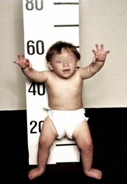
Lymphangioma patients often appear healthy. Physical examination of patients with lymphangioma is usually remarkable for painless, compressible, soft neck mass that often transilluminates is a diagnostic finding on physical exam.
Physical Examination
Physical examination of patients with lymphangioma is usually normal except cystic swellings found in the residing organs.[1]
Appearance of the Patient
- Patients with lymphangioma usually appear normal.
Vital Signs
- No fever
- Normal Heart rate with regular pulse
- Normal respiration rate
- Within normal range
Skin
- Skin examination of patients with lymphangioma is usually normal or the skin overlying cystic swelling may look bluish.
-
 Description (Adapted from Dermatology Atlas)

HEENT
- HEENT examination of patients with lymphangioma is usually normal.
- No Abnormalities of the head/hair
- No Evidence of trauma
- No Icteric sclera
- No Nystagmus
- Extra-ocular movements may be abnormal depending on retro-orbital location of lymphangioma
- Pupils reactive to light / reactive to accommodation
- Hearing acuity is normal
- Weber test is normal
- Rinne test is normal
- No Exudate from the ear canal
- No Tenderness upon palpation of the ear pinnae/tragus (anterior to ear canal)
- No Inflamed nares / congested nares
- No Purulent exudate from the nares
- No Facial tenderness
- No Erythematous throat with/without tonsillar swelling, exudates, and/or petechiae
Neck
- Neck examination of patients with lymphangioma may show soft cystic swelling.
- No Jugular venous distension
- No Carotid bruits may be auscultated unilaterally/bilaterally using the bell/diaphragm of the otoscope
- No Lymphadenopathy (describe location, size, tenderness, mobility, and symmetry)
- No Thyromegaly / thyroid nodules
- No Hepatojugular reflux
Lungs
- Asymmetric chest expansion OR decreased chest expansion depending on location of lymphangioma
- Lungs are hyporesonant.
- No Fine/coarse crackles upon auscultation of the lung bases/apices unilaterally/bilaterally
- No Rhonchi
- Vesicular breath sounds OR distant breath sounds may be heard.
- Wheezing may be present
- Egophony absent
- Bronchophony present
- Reduced tactile fremitus
Heart
- Cardiovascular examination of patients with lymphangioma is usually normal.
Abdomen
- Abdominal examination of patients with lymphangioma is usually normal.
Back
- Back examination of patients with lymphangioma is usually normal.
Genitourinary
- Genitourinary examination of patients with lymphangioma is usually normal.
Neuromuscular
- Neuromuscular examination of patients with lymphangioma is usually normal.
Extremities
- Extremities examination of patients with lymphangioma is usually normal.
References
- ↑ Patoulias D, Patoulias I, Kaselas C, Kalogirou M, Kyriakos C, Konstantinos F, Feidantsis T, Eleni P (2017). "Cystic Lymphangioma of the Chest Wall in a 5-Year-Old Male Patient: A Rare and Atypical Localization-A Case Report and Comprehensive Review of the Literature". Case Rep Pediatr. 2017: 2083204. doi:10.1155/2017/2083204. PMC 5672607. PMID 29201481.