Hurler syndrome physical examination
|
Hurler Syndrome Microchapters |
|
Diagnosis |
|---|
|
Treatment |
|
Case Studies |
|
Hurler syndrome physical examination On the Web |
|
American Roentgen Ray Society Images of Hurler syndrome physical examination |
|
Risk calculators and risk factors for Hurler syndrome physical examination |
Editor-In-Chief: C. Michael Gibson, M.S., M.D. [1]; Associate Editor(s)-in-Chief: Jesus Rosario Hernandez, M.D. [2].
Please help WikiDoc by adding content here. It's easy! Click here to learn about editing.
Overview
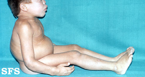
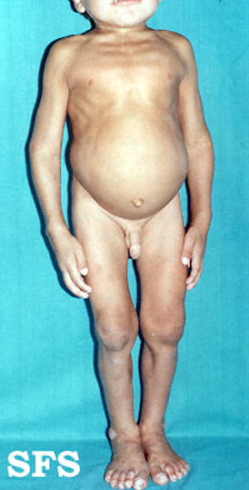
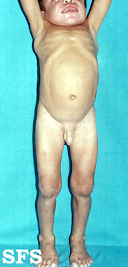
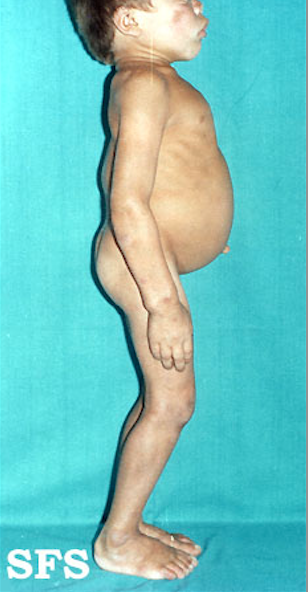
The condition is marked by progressive deterioration, hepatosplenomegaly, dwarfism and gargoyle-like faces. There is a progressive mental retardation, with death frequently occurring by the age of 10 years.
Developmental delay is evident by the end of the first year, and patients usually stop developing between ages 2 and 4. This is followed by progressive mental decline and loss of physical skills. Language may be limited due to hearing loss and an enlarged tongue. In time, the clear layers of the cornea become clouded and retinas may begin to degenerate. Carpal tunnel syndrome (or similar compression of nerves elsewhere in the body) and restricted joint movement are common.
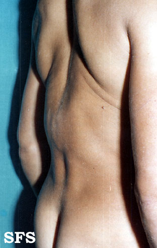
Physical examination
Gallery
Head


Trunk
-
 Hurler syndrome.
 -
-
-
-