Hepatocyte
Editor-In-Chief: C. Michael Gibson, M.S., M.D. [1]


Hepatocytes make up 70-80% of the cytoplasmic mass of the liver. These cells are involved in protein synthesis, protein storage and transformation of carbohydrates, synthesis of cholesterol, bile salts and phospholipids, and detoxification, modification and excretion of exogenous and endogenous substances.
The hepatocyte also initiates the formation and secretion of bile.
Hepatocyte histology
Hepatocytes display an eosinophilic cytoplasm, reflecting numerous mitochondria, and basophilic stippling due to large amounts of rough endoplasmic reticulum and free ribosomes. Brown lipofuscin granules are also observed (with increasing age) together with irregular unstained areas of cytoplasm; these correspond to cytoplasmic glycogen and lipid stores removed during histological preparation. The average life span of the hepatocyte is 5 months; they are able to regenerate.
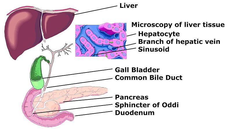
Hepatocyte nuclei are round with dispersed chromatin and prominent nucleoli. Anisokaryosis is common and reflects tetraploidy & polyploidy, a normal feature of over 50% of hepatocytes. Binucleate cells are also common. Hepatocytes are organised into plates separated by vascular channels (sinusoids), an arrangement supported by a reticulin (collagen type III) network. The hepatocyte plates are one cell thick in mammals and two cells thick in the chicken. Sinusoids display a discontinuous, fenestrated endothelial cell lining. The endothelial cells have no basement membrane and are separated from the hepatocytes by the space of Disse which drains lymph into the portal tract lymphatics. Kupffer cells are scattered between endothelial cells; they are part of the reticuloendothelial system and phagocytose spent erythrocytes. Stellate (Ito) cells store vitamin A and produce extracellular matrix and collagen; they are also distributed amongst endothelial cells but are difficult to visualise by light microscopy.
Hepatocytes are an important physiological example for evalutation of both biological and metabolic effects of xenobiotics. They do not proliferate in culture. Hepatocytes are intensely sensitive to damage during the cycles of cryopreservation including freezing and thawing. Even after the addition of classical cryoprotectants there is still damage done while being cryopreserved. [1]
Protein synthesis
The hepatocyte is a cell in the body that manufactures serum albumin, fibrinogen, and the prothrombin group of clotting factors. It is the main site for the synthesis of lipoproteins, ceruloplasmin, transferrin, complement and glycoproteins. Hepatocytes manufacture their own structural proteins and intracellular enzymes.
Synthesis of proteins is undertaken by the rough endoplasmic reticulum (RER), and both the rough and smooth endoplasmic reticulum (SER) are involved in secretion of the proteins formed. The endoplasmic reticulum (ER) is involved in conjugation of proteins to lipid and carbohydrate moieties synthesized by, or modified within, the hepatocytes.
Carbohydrate metabolism
The liver forms fatty acids from carbohydrates and synthesizes triglycerides from fatty acids and glycerol. Hepatocytes also synthesize apoproteins with which they then assemble and export lipoproteins (VLDL, HDL).
Lipid metabolism
The liver receives many lipids from the systemic circulation and metabolizes chylomicron remnants. It also synthesizes cholesterol from acetate and then further synthesizes bile salts. The liver is the sole site of formation of bile salts.
Detoxification
Hepatocytes have the ability to metabolize, detoxify, and inactivate exogenous compounds such as drugs and insecticides, and endogenous compounds such as steroids.
The drainage of the intestinal venous blood into the liver requires efficient detoxification of miscellaneous absorbed substances to maintain homeostasis and protect the body against ingested toxins.
One of the detoxifying functions of hepatocytes is to modify ammonia into urea for excretion.
Additional images
-
 Schemic diagram of Biliary system

References
- ↑ Hamel et al; "Wheat Extracts as an Efficient Cryoprotective Agent for Primary Cultures of Rat Hepatocytes": published online 21 Aug 2006 in Wiley Interscience www.interscience.wiley.com. Department des sciences bogiques, Montreal University.
External links
- Histology image: 22101ooa – Histology Learning System at Boston University - "Ultrastructure of the Cell: hepatocytes and sinusoids"
- Hepatic Histology: Hepatocytes (Colorado State University
cs:Hepatocyt de:Hepatozyt it:Epatocita lt:Hepatocitas mk:Хепатоцит fi:Maksasolu sv:Hepatocyt