Hamman-Rich syndrome pathophysiology
|
Hamman-Rich syndrome Microchapters |
|
Diagnosis |
|---|
|
Treatment |
|
Case Studies |
|
Hamman-Rich syndrome pathophysiology On the Web |
|
American Roentgen Ray Society Images of Hamman-Rich syndrome pathophysiology |
|
Risk calculators and risk factors for Hamman-Rich syndrome pathophysiology |
Editor-In-Chief: C. Michael Gibson, M.S., M.D. [1]; Associate Editor(s)-in-Chief: Chandrakala Yannam, MD [2]
Overview
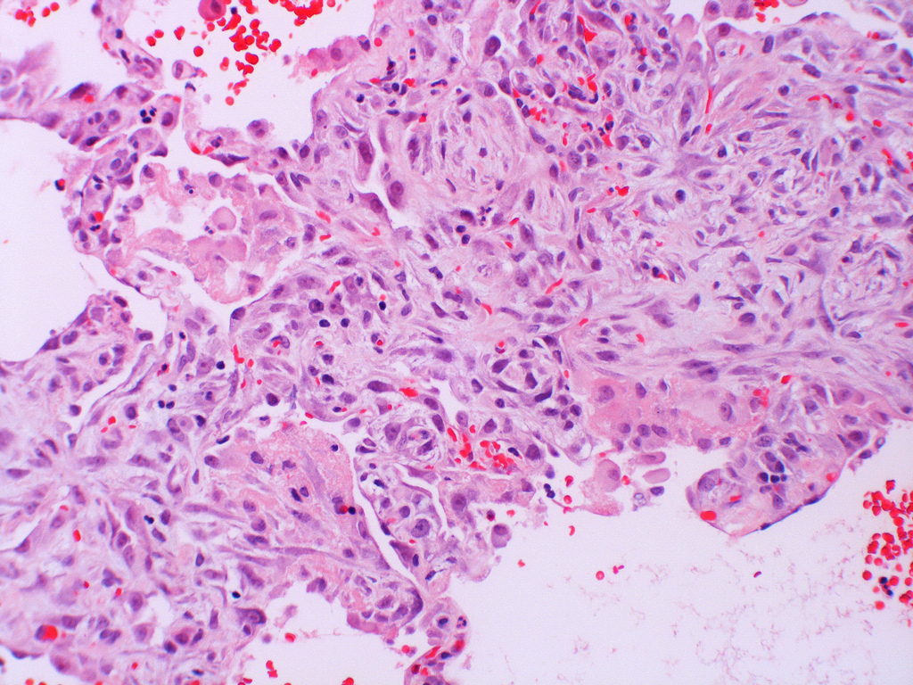
Acute interstitial pneumonitis shows the histopathologic appearance of diffuse alveolar damage. On gross examination, lungs appear firm, heavy and have a dark red or beefy appearance and show irregular areas of consolidation and fibrosis. On microscopic examination, acute interstitial pneumonitis shows bilateral, temporal uniformity of the diffuse alveolar damage, hyaline membrane deposition and extensive fibroblastic and myofibroblastic proliferation.
Pathophysiology
Pathogenesis
- The mechanism of initial injury to the pulmonary epithelium in acute interstitial pneumonitis is not known.
- Based on the findings of lung biopsy, the uniformity and extent of the injury, it is assumed that a single insult triggers the pathologic process.
- On microscopy, appearance of hyaline membrane remenats and organizing alveolar exudate may suggest that acute interstitial pnemonia progresses from the exudative stage of diffuse alveolar damage.
- Diffuse alveolar damage occurs in 3 stages.[1][2]
- The rapid and abrupt onset of a widespread injury pattern may be suggested as an initiating factor involving pulmonary endothelium. This is followed by damage to alveolar epithelium and cell death, that leads to release of mediating factors such as tumor necrosis factor alpha, interleukins and monocyte chemoattractant factor.
- Followed by the influx of neutrophils into the alveolar spaces and alveolar walls leads to further cellular damage, by the release of toxic oxygen free radicals and proteases.
- Neutrophils and other inflammatory cells can contribute to the progression of the epithelial cell injury and airspace exudation.
- The extent of epithelial injury and basement membrane damage may modulate the nature and extent of the subsequent fibroblastic response in acute interstitial pneumonitis.
- Following the acute phase, a stage of organization occurs.
- The hyaline membranes are resorbed into the alveolar septa and overgrown by proliferating type II pneumocytes.
- In fibrotic phase, proliferation of fibroblasts and differentiation into myofibroblasts leads to the production of collagen, causes widening of the alveolar septa, and organization of the alveolar exudate.
- Collapse of alveolar wall and apposition, associated with reepithelization of the fibrotic exudate within the alveolar space may contribute to the severity and extent of the fibrotic process.
Gross Pathology
- Patients with acute interstitial pneumonitis, gross appearance of lungs is identical to the patients with ARDS.[3]
- The gross appearance of lungs correlates with the stage of the diffuse alveolar damage.
- In the early phase, the lungs are firm, boggy, and have a dark red or beefy appearance.
- In later phases, the lungs are extremely heavy due to edema and show irregular areas of dense consolidation and fibrosis.
- As the fibrosis progresses, cobblestoning of the pleural surface may occur.
- Formation of peripheral cysts and honeycombing may suggest the possibility of underlying chronic fibrotic lung disease.
Microscopic Pathology
- On microscopic histopathological analysis, Acute interstitial pneumonitis will show following features:[4][5]
- Diffuse alveolar damage
- Hyaline membrane formation
- Interstitial fibrosis:
- It is diffuse, bilateral, uniform temporally with extensive fibroblastic and myofibroblastic proliferation and relatively less collagen deposition.
- The uniformity of the fibroblastic/myofibroblastic proliferation and prominent activity distinguish AIP from the other types of idiopathic interstitial pneumonia.
- Type II pneumocyte hyperplasia with cytologic atypia and bronchiolar squamous metaplasia is present.
- Thickening and distortion of alveolar septa caused by spindle cell proliferation.
- Intraluminal polypoid plugs formation
- Organizing thrombi in small and medium-sized arteries
- Biopsies taken in the later course of the disease show:
- Enlarged and remodeled airspaces that resemble honeycomb change of UIP (usual interstitial fibrosis), but extensive fibroblastic/ myofibroblastic proliferation and collagen deposition is still present within the walls of the alveoli.

References
- ↑ Nur Urer H, Ersoy G, Yılmazbayhan ED (2012). "Diffuse alveolar damage of the lungs in forensic autopsies: assessment of histopathological stages and causes of death". ScientificWorldJournal. 2012: 657316. doi:10.1100/2012/657316. PMC 3458269. PMID 23028252.
- ↑ Kang D, Nakayama T, Togashi M, Yamamoto M, Takahashi M, Kunugi S, Ishizaki M, Fukuda Y (November 2009). "Two forms of diffuse alveolar damage in the lungs of patients with acute respiratory distress syndrome". Hum. Pathol. 40 (11): 1618–27. doi:10.1016/j.humpath.2009.04.019. PMID 19647854.
- ↑ Tomashefski JF (September 2000). "Pulmonary pathology of acute respiratory distress syndrome". Clin. Chest Med. 21 (3): 435–66. PMID 11019719.
- ↑ Mukhopadhyay S, Parambil JG (October 2012). "Acute interstitial pneumonia (AIP): relationship to Hamman-Rich syndrome, diffuse alveolar damage (DAD), and acute respiratory distress syndrome (ARDS)". Semin Respir Crit Care Med. 33 (5): 476–85. doi:10.1055/s-0032-1325158. PMID 23001802.
- ↑ Bonaccorsi A, Cancellieri A, Chilosi M, Trisolini R, Boaron M, Crimi N, Poletti V (January 2003). "Acute interstitial pneumonia: report of a series". Eur. Respir. J. 21 (1): 187–91. PMID 12570127.
- ↑ By Yale Rosen [CC BY-SA 2.0 (https://creativecommons.org/licenses/by-sa/2.0)], via Wikimedia Commons