Dilated cardiomyopathy electrocardiogram
|
Dilated cardiomyopathy Microchapters |
|
Diagnosis |
|---|
|
Treatment |
|
Case Studies |
|
Dilated cardiomyopathy electrocardiogram On the Web |
|
American Roentgen Ray Society Images of Dilated cardiomyopathy electrocardiogram |
|
Risk calculators and risk factors for Dilated cardiomyopathy electrocardiogram |
Editor-In-Chief: C. Michael Gibson, M.S., M.D. [1]; Associate Editor(s)-in-Chief: Abdelrahman Ibrahim Abushouk, MD[2]
Overview
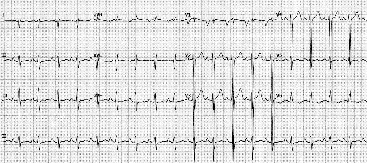
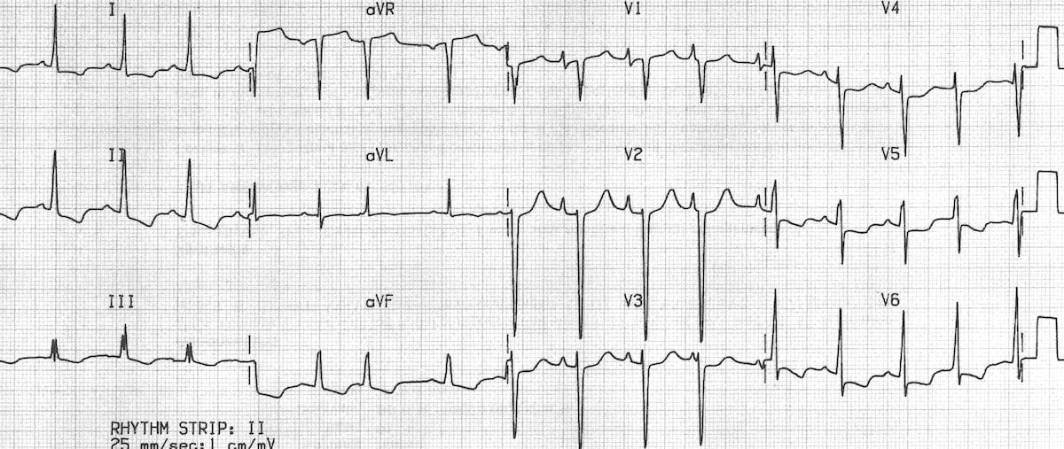
ECG may show evidence of left ventricular hypertrophy, atrial fibrillation or premature ventricular complexes, or conduction delays, AV nodal block, or left bundle branch block may be observed.
Electrocardiogram
ECG may show evidence of the following findings:
- Left ventricular hypertrophy
- Atrial fibrillation or premature ventricular complexes
- Conduction delays, AV nodal block, or left bundle branch block may be observed.
Some studies have shown that left ventricular hypertrophy, altered heart rate, and anterolateral T-wave inversion can predict the risk of mortality or heart transplantation in dilated cardiomyopathy patients.[1][2]


References
- ↑ Merlo M, Zaffalon D, Stolfo D, Altinier A, Barbati G, Zecchin M; et al. (2019). "ECG in dilated cardiomyopathy: specific findings and long-term prognostic significance". J Cardiovasc Med (Hagerstown). 20 (7): 450–458. doi:10.2459/JCM.0000000000000804. PMID 30985353.
- ↑ Momiyama Y, Mitamura H, Kimura M (1994). "ECG characteristics of dilated cardiomyopathy". J Electrocardiol. 27 (4): 323–8. doi:10.1016/s0022-0736(05)80270-5. PMID 7815010.
- ↑ https://litfl.com/wp-content/uploads/2018/08/ECG-Ischaemic-dilated-cardiomyopathy-1.jpg
- ↑ https://litfl.com/wp-content/uploads/2018/08/ECG-Idiopathic-dilated-cardiomyopathy-Biatrial-hypertrophy.jpg