Amblyopia
| Amblyopia | |
 | |
|---|---|
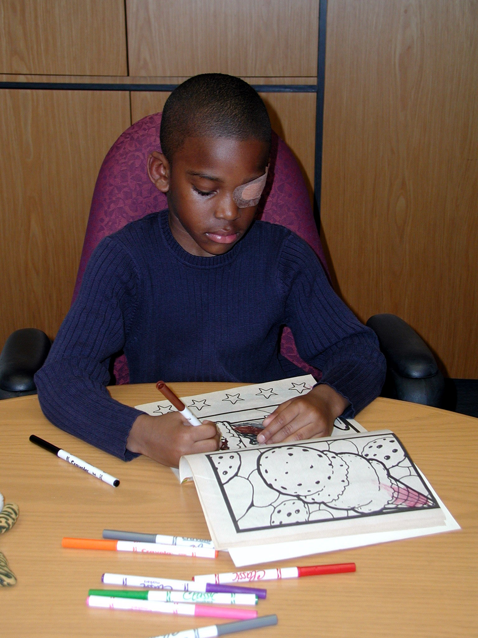
| A child wearing an adhesive eyepatch to correct amblyopia | |
| ICD-10 | H53.0 |
| ICD-9 | 368.0 |
| DiseasesDB | 503 |
| MedlinePlus | 001014 |
| MeSH | D000550 |
For patient information click here
|
WikiDoc Resources for Amblyopia |
|
Articles |
|---|
|
Most recent articles on Amblyopia |
|
Media |
|
Evidence Based Medicine |
|
Clinical Trials |
|
Ongoing Trials on Amblyopia at Clinical Trials.gov Clinical Trials on Amblyopia at Google
|
|
Guidelines / Policies / Govt |
|
US National Guidelines Clearinghouse on Amblyopia
|
|
Books |
|
News |
|
Commentary |
|
Definitions |
|
Patient Resources / Community |
|
Patient resources on Amblyopia Discussion groups on Amblyopia Directions to Hospitals Treating Amblyopia Risk calculators and risk factors for Amblyopia
|
|
Healthcare Provider Resources |
|
Causes & Risk Factors for Amblyopia |
|
Continuing Medical Education (CME) |
|
International |
|
|
|
Business |
|
Experimental / Informatics |
Editor-In-Chief: C. Michael Gibson, M.S., M.D. [1]
Overview
Amblyopia, or lazy eye, is a disorder of the eye that is characterized by poor or indistinct vision in an eye that is otherwise physically normal, or out of proportion to associated structural abnormalities. It has been estimated to affect 1–5% of the population.[1]
The problem is caused by either no transmission or poor transmission of the visual image to the brain for a sustained period of dysfunction or during early childhood. Amblyopia normally only affects one eye, but it is possible to be amblyopic in both eyes if both are similarly deprived of a good, clear visual image. Detecting the condition in early childhood increases the chance of successful treatment.
Physiology
Amblyopia is a developmental problem in the brain, not an organic problem in the eye (although organic problems can induce amblyopic symptoms which persist after the organic problem has resolved).[2] The part of the brain corresponding to the visual system from the affected eye is not stimulated properly, and develops abnormally. This has been confirmed via direct brain examination. David H. Hubel and Torsten Wiesel won the Nobel Prize in Physiology or Medicine in 1981 for their work demonstrating the irreversible damage to ocular dominance columns produced in kittens by sufficient visual deprivation during the so-called "critical period". The maximum critical period in humans is from birth to two years old.[2]
Symptoms
Many amblyopes, especially those who are only mildly so, are not even aware they have the condition until tested at older ages, since the vision in their stronger eye is normal. However, people who have severe amblyopia may experience associated visual disorders, most notably poor depth perception. Amblyopes suffer from poor spatial acuity, low sensitivity to contrast and some "higher-level" deficits to vision such as reduced sensitivity to motion.[3] These deficits are usually specific to the amblyopic eye, not the unaffected "fellow" eye. Amblyopes also suffer from problems of binocular vision such as limited stereoscopic depth perception and usually have difficulty seeing the three-dimensional images in hidden stereoscopic displays such as autostereograms.[4] However perception of depth from monocular cues such as size, perspective, and motion parallax is normal.
Types
Amblyopia can be caused by deprivation of vision early in life, by strabismus (misaligned eyes), by vision-obstructing disorders, or by anisometropia (different degrees of myopia or hyperopia in each eye).
Strabismic amblyopia
Strabismus, sometimes erroneously also called lazy eye, is a condition in which the eyes are misaligned in a variety of different ways. Strabismus usually results in normal vision in the preferred sighting eye, but may cause abnormal vision in the deviating or strabismic eye due to the discrepancy between the images projecting to the brain from the two eyes.[5] Adult-onset strabismus usually causes double vision (diplopia), since the two eyes are not fixated on the same object. Children's brains, however, are more plastic, and therefore can more easily adapt by ignoring images from one of the eyes, eliminating the double vision (suppression (eye)). This plastic response of the brain, however, interrupts the brain's normal development, resulting in the amblyopia.
Strabismic amblyopia is treated by clarifying the visual image with glasses, and/or encouraging use of the amblyopic eye with patching or pharmacologic penalization (usually by applying atropine drops to the dominant eye to temporarily paralyze the muscles and weaken vision in the good eye—this helps to prevent the bullying and teasing associated with wearing a patch). The ocular alignment itself may be treated with surgical or non-surgical methods, depending on the type and severity of the strabismus.[6]
Refractive or anisometropic amblyopia
Refractive amblyopia may result from anisometropia (unequal refractive errors between the two eyes). Anisometropia exists when there is a difference in the refraction between the two eyes. The eye with less far-sighted (hyperopic) refractive error provides the brain with a clearer image, and is favored by the brain. Refractive amblyopia is usually less severe than strabismic amblyopia and is commonly missed by primary care physicians because of its less dramatic appearance and lack of obvious physical manifestation, such as with strabismus.[7]
Frequently, amblyopia is associated with a combination of anisometropia and strabismus.
Pure refractive amblyopia is treated by correcting the refractive error early with prescription lenses. Vision therapy and/or eye patching can also be used to develop and/or improve visual abilities, binocular vision, depth perception, etc.
Meridional amblyopia is a mild condition in which lines are seen less clearly at some orientations than others after full refractive correction. An individual who had an astigmatism at a young age that was not corrected by glasses will later have astigmatism that cannot be optically corrected.
Form-deprivation and occlusion amblyopia
Form-deprivation amblyopia (Amblyopia ex anopsia) results when the ocular media become opaque, such as is the case with cataracts or corneal scarring from forceps injuries during birth.[8] These opacities prevent adequate sensory input from reaching the eye, and therefore disrupt visual development. If not treated in a timely fashion, amblyopia may persist even after the cause of the opacity is removed. Sometimes, drooping of the eyelid (ptosis) or some other problem causes the upper eyelid to physically occlude a child's vision, which may cause amblyopia quickly. Occlusion amblyopia may be a complication of a hemangioma that blocks some or all of the eye.
Treatment and prognosis
Treatment of strabismic or anisometropic amblyopia consists of correcting the optical deficit and forcing use of the amblyopic eye, either by patching the good eye, or by instilling topical atropine in the eye with better vision. One should also be wary of over-patching or over-penalizing the good eye when treating for amblyopia, as this can create so-called "reverse amblyopia" in the other eye.[6][9]
Form deprivation amblyopia is treated by removing the opacity as soon as possible followed by patching or penalizing the good eye to encourage use of the amblyopic eye.[6]
Although the best outcome is achieved if treatment is started before age 5, research has shown that children older than age 10 and some adults can show improvement in the affected eye. Children from 7 to 12 who wore an eye patch and performed near point activities (vision therapy) were four times as likely to show a two line improvement on a standard 11 line eye chart than amblyopic children who did not receive treatment. Children 13 to 17 showed improvement as well, albeit in smaller amounts than younger children. (NEI-funded Pediatric Eye Disease Investigator Group, 2005)[6][10] Some claim the controversial[11] Bates Method can reverse amblyopia, however, this assertion is unfounded. [12]
See also
References
- ↑ Weber, JL; Wood, Joanne (2005). "Amblyopia: Prevalence, Natural History, Functional Effects and Treatment" (PDF). Clinical and Experimental Optometry. 88 (6): 365–375.
- ↑ McKee, SP., Levi, DM., Movshon, JA. (2003). "The pattern of visual deficits in amblyopia" (PDF). J Vision. 3 (5): 380–405.
- ↑ Hess, R.F., Mansouri, B., Dakin, S.C., & Allen, H.A. (2006). "Integration of local motion is normal in amblyopia". J Opt Soc Am A Opt Image Sci Vis. 23 (5): 986–992.
- ↑ Tyler, C.W. (2004). "Binocular Vision In, Duane's Foundations of Clinical Ophthalmology. Vol. 2, Tasman W., Jaeger E.A. (Eds.), J.B. Lippincott Co.: Philadelphia".
- ↑ Levi, D.M. (2006). "Visual processing in amblyopia: human studies". Strabismus. 14 (1): 11–19.
- ↑ 6.0 6.1 6.2 6.3 Holmes, Repka, Kraker & Clarke (2006). "The treatment of amblyopia". Strabismus. 14 (1): 37–42.
- ↑ http://www.aafp.org/afp/20010815/623.html
- ↑ http://archopht.ama-assn.org/cgi/content/abstract/99/12/2137
- ↑ http://www.nei.nih.gov/health/amblyopia/index.asp
- ↑ Pediatric Eye Disease Investigator Group (2005). "Randomized trial of treatment of amblyopia in children aged 7 to 17 years". Archives of Ophthalmology. 123 (April): 437–447.
- ↑ Robyn E. Bradley (September 23, 2003). "Advocates see only benefits from eye exercises" (PDF). The Boston Globe (MA).
- ↑ Rawstron JA, Burley CD, Elder MJ (2005). "A systematic review of the applicability and efficacy of eye exercises". J Pediatr Ophthalmol Strabismus. 42 (2): 82–8.
External links
- Amblyopia Resource Guide from the National Eye Institute (NEI).
- The Lazy Eye Site - enhancing awareness of squint/lazy eye treatment available
- http://www.aapos.org/displaycommon.cfm?an=1&subarticlenbr=63]
- eMedicine - Amblyopia
- All About Amblyopia - FAQ, Recent Research, etc.
- Convergence Insufficiency - Common Visual Condition Confused with Lazy Eye
- BBC article on new study that finds lazy eye can be treated in teen years
- Prevent Blindness America Amblyopia FAQ
- Strabismus - Lazy Eye and Strabismus are Not Same Condition
- The Amblyopia Foundation of America
- U.S. National Institutes of Health - Older Children Can Benefit from Treatment
- Treatment using VR software being trialled (Eastgate, et.al. Nottingham University)
- Interactive Binocular Treatment for Amblyopia