Adipose

In anatomy, adipose tissue or fat is loose connective tissue composed of adipocytes. Its main role is to store energy in the form of fat, although it also cushions and insulates the body. Obesity in humans and most animals is not dependent on the amount of body weight, but on the amount of body fat—specifically adipose tissue. Two types of adipose tissue exist: white adipose tissue (WAT) and brown adipose tissue (BAT). Adipose tissue also serves as an important endocrine organ[1] by producing recently-discovered hormones such as leptin, resistin and the cytokine TNFα. The formation of adipose tissue appears to be controlled by the adipose gene.
Anatomical features


In humans, adipose tissue is located beneath the skin and is also found around internal organs. Adipose tissue is found in specific locations, which are referred to as 'adipose depots'. Adipose tissue contains several cell types, with the highest percentage of cells being adipocytes, which contain fat droplets. Other cell types include fibroblasts, macrophages and endothelial cells. Adipose tissue contains many small blood vessels. In the integumentary system, which includes the skin, it accumulates in the deepest level, the subcutaneous layer, providing insulation from heat and cold. Around organs, it provides protective padding. However, its main function is to be a reserve of lipids, which can be burned to meet the energy needs of the body. Adipose depots in different parts of the body have different biochemical profiles.
In a severely obese person, excess adipose tissue hanging downward from the abdomen is referred to as a panniculus (or pannus). A panniculus complicates surgery of the morbidly obese. The panniculus may remain as a literal "apron of skin" if a severely obese person quickly loses large amounts of weight (a common result of gastric bypass surgery). This condition cannot easily be corrected through diet and exercise, as the panniculus consists of adipocytes and other supporting cell types shrunken to their minimum volume and diameter. Reconstructive surgery is one way to fix the problem.
In mice, there are eight major adipose depots, four of which are within the abdominal cavity: the paired gonadal depots are attached to the uterus and ovaries in females and the epididymis and testes in males, the paired retroperitoneal depots are found along the dorsal wall of the abdomen, surrounding the kidney, and when massive extend into the pelvis. The mesenteric depot forms a glue-like web that supports the intestines, and the omental depot, which originates near the stomach and spleen and when massive extends into the ventral abdomen. Both the mesenteric and omental depots incorporate much lymphoid tissue as lymph nodes and milky spots respectively. The two superficial depots are the paired inguinal depots, which are found anterior to the upper segment of the hind limbs (underneath the skin) and the subscapular depots, paired medial mixtures of brown adipose tissue adjacent to regions of white adipose tissue, which are found under the skin between the dorsal crests of the scapulae. The layer of brown adipose tissue in this depot is often covered by a “frosting” of white adipose tissue, sometimes these two types of fat (brown and white) are hard to distinguish. The inguinal depots enclose the inguinal group of lymph nodes. Minor depots include the pericardial which surrounds the heart, and the paired popliteal depots, between the major muscles behind the knees, each containing one large lymph node(Pond 1998). Of all the depots in the mouse, the gonadal depots are the largest and the most easily dissected (Cinti, 1999), comprising about 30% of dissectible fat, e.g., (Bachmanov et al. 2001).
Physiology
This article needs additional citations for verification. (July 2007) (Learn how and when to remove this template message) |
Free fatty acid is "liberated" from lipoproteins by lipoprotein lipase (LPL) and enters the adipocyte, where it is reassembled into triglycerides by esterising it onto glycerol. Human fat tissue contains about 87% lipids.
In humans, lipolysis is controlled though the balanced control of lipolytic B-adrenergic receptors and a2A-andronergic receptor mediated antilipolysis.
Fat is not laid down when there is a surplus available and stored passively until it is needed; rather it is constantly being stored in and released from each cell.
Fat cells have an important physiological role in maintaining triglyceride and free fatty acid levels, as well as determining insulin resistance. Abdominal fat has a different metabolic profile—being more prone to induce insulin resistance. This explains to a large degree why central obesity is a marker of impaired glucose tolerance and is an independent risk factor for cardiovascular disease (even in the absence of diabetes mellitus and hypertension).
Recent advances in biotechnology have allowed for the harvesting of adult stem cells from adipose tissue, allowing stimulation of tissue regrowth using a patient's own cells. The use of a patient's own cells reduces the chance of tissue rejection and avoids the ethical issues associated with the use of human embryonic stem cells.
Adipose tissue is the greatest peripheral source of aromatase in both males and females contributing to the production of estradiol.
Hormones secreted by adipose tissue include:
- Adiponectin
- Resistin
- Angiotensin
- Plasminogen activator inhibitor-1 (PAI-1)
- TNFα
- IL-6
- Leptin
- Estradiol (E2)
Adipose tissues also secret a type of cytokines (cell-to-cell signalling proteins) called adipokines (adipocytokines) which play a role in obesity-associated complications.
Brown fat
A specialised form of adipose tissue in human infants, and some animals, is brown fat or brown adipose tissue. It is located mainly around the neck and large blood vessels of the thorax. This specialised tissue can generate heat by "uncoupling" the respiratory chain of oxidative phosphorylation within mitochondria, leading to the breakdown of fatty acids. This thermogenic process may be vital in neonates exposed to the cold, who then require this thermogenesis to keep warm as they are unable to shiver, or take other actions to keep themselves warm.[2]
Attempts to stimulate this process pharmacologically have so far been unsuccessful, but might in the future be a target of weight loss therapy.
Genetics
In 2007, researchers isolated the adipose gene, which apparently serves to keep animals lean during times of plenty. Increased adipose gene activity was associated with slimmer individuals.[3]
Cultural and social role
Excess adipose tissue on a human can lead to medical problems; however, a round or large figure does not of itself imply a medical problem, and is sometimes not primarily caused by adipose tissue. For a discussion of the aesthetic and medical significance of body shape, see dieting and obesity.
Additional images
-
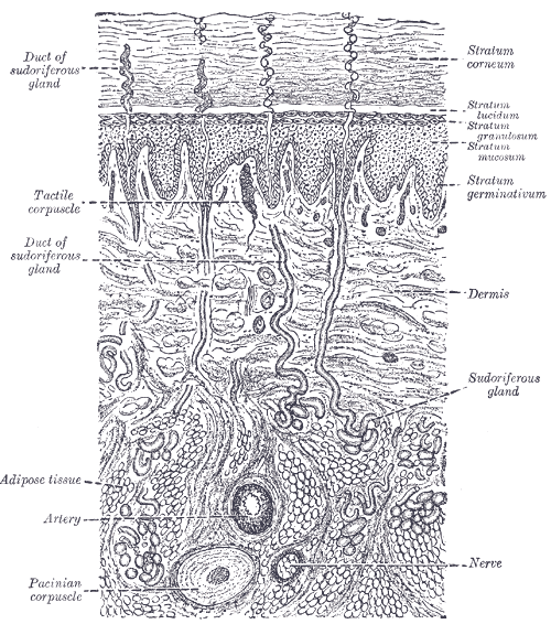 diagrammatic sectional view of the skin (magnified).
 -
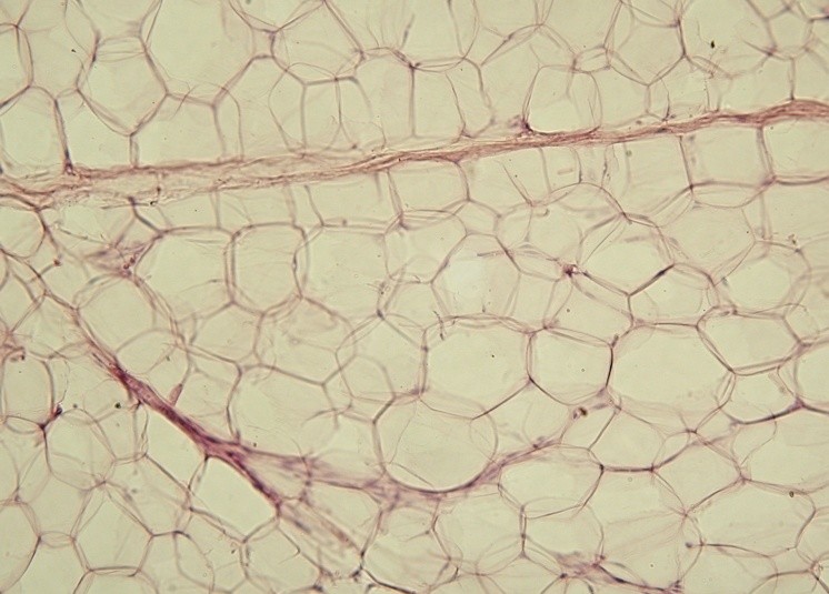 Yellow adipose tissue in paraffin section


References
- ↑ Kershaw EE, Flier JS (2004). "Adipose tissue as an endocrine organ". J. Clin. Endocrinol. Metab. 89 (6): 2548–56. doi:10.1210/jc.2004-0395. PMID 15181022.
- ↑ Himms-Hagen, J. (August 1990) "Brown adipose tissue thermogenesis: interdisciplinary studies" The FASEB Journal (Federation of American Societies for Experimental Biology) 4(11): pp. 2890-2898
- ↑ Suh, Jae Myoung et al. (September 2007) "Adipose Is a Conserved Dosage-Sensitive Antiobesity Gene" Cell Metabolism 6(3): pp. 195-207
- Bachmanov AA, Reed DR, Tordoff MG, Price RA, Beauchamp GK (2001) Nutrient preference and diet-induced adiposity in C57BL/6ByJ and 129P3/J mice. Physiology and Behavior 72, 603-613
- Cinti S (1999) The adipose organ. Editrice Kurtis, Milano
- Pond CM (1998) The fats of life. Cambridge University Press, Cambridge
See also
- Bioelectrical impedance analysis: a method to measure body fat percentage.
- Body fat percentage
- Body fat meter
- Cellulite
- Obesity
- Stem Cells
- Apelin
It has been suggested that Subcutaneous fat be merged into this article. (Discuss) Proposed since January 2007. |
It has been suggested that Visceral fat be merged into this article. (Discuss) Proposed since January 2007. |
ca:Teixit adipós de:Fettgewebe el:Λιπώδης ιστός gl:Tecido adiposo it:Tessuto adiposo lt:Riebalinis audinys nl:Vetweefsel fi:Rasvakudos sv:Fettvävnad