Papillary thyroid cancer diagnostic study of choice: Difference between revisions
(Created page with "__NOTOC__ {{Papillary thyroid cancer}} {{CMG}}; {{AE}} {{Ammu}} ==Overview== According to the American Joint Committee on Cancer (AJCC)<ref> Stage Information for Thyroid Canc...") |
No edit summary |
||
| Line 10: | Line 10: | ||
===Staging=== | ===Staging=== | ||
Based on overall [[cancer staging]] into stages I to IV, papillary thyroid cancer has a [[5-year survival rate]] of 100 percent for stages I and II, 93 percent for stage III and 51 percent for stage IV.<ref name=acc>[http://www.cancer.org/cancer/thyroidcancer/detailedguide/thyroid-cancer-survival-rates cancer.org > Thyroid Cancer] By the American Cancer Society. In turn citing: AJCC Cancer Staging Manual (7th ed).</ref> | Based on overall [[cancer staging]] into stages I to IV, papillary thyroid cancer has a [[5-year survival rate]] of 100 percent for stages I and II, 93 percent for stage III and 51 percent for stage IV.<ref name=acc>[http://www.cancer.org/cancer/thyroidcancer/detailedguide/thyroid-cancer-survival-rates cancer.org > Thyroid Cancer] By the American Cancer Society. In turn citing: AJCC Cancer Staging Manual (7th ed).</ref> | ||
====Papillary thyroid cancer in patients younger than 45 years==== | ====Papillary thyroid cancer in patients younger than 45 years==== | ||
| Line 232: | Line 231: | ||
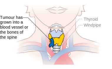
File:Diagram showing stage T4b thyroid cancer CRUK 273.png|Stage T4b thyroid cancer | File:Diagram showing stage T4b thyroid cancer CRUK 273.png|Stage T4b thyroid cancer | ||
</gallery> | </gallery> | ||
==Key Biopsy Findings in Follicular Thyroid Cancer== | |||
*Characteristic [[Orphan Annie eye]] nuclear inclusions (nuclei with uniform [[staining]], which appear empty)<ref>{{cite web|url=http://esynopsis.uchc.edu/eAtlas/Endo/1802.htm|title=Papillary Carcinoma of Thyroid (Hi Pow)|publisher=University of Connecticut Health Center|accessdate=2008-09-14}}</ref> and [[psammoma]] bodies on light microscopy. The former is useful in identifying the follicular variant of papillary thyroid carcinomas.<ref name="pmid11442006">{{cite journal |author=Yang GC, Liebeskind D, Messina AV |title=Ultrasound-guided fine-needle aspiration of the thyroid assessed by Ultrafast Papanicolaou stain: data from 1135 biopsies with a two- to six-year follow-up |journal=Thyroid |volume=11 |issue=6 |pages=581–89 |year=2001 |pmid=11442006 |doi=10.1089/105072501750302895}}</ref> | |||
* Histologically papillary carcinoma demonstrates 'delicate stalks of epithelial cells' which account for its name. | |||
==Reference== | ==Reference== | ||
{{Reflist|2}} | {{Reflist|2}} | ||
Revision as of 20:00, 5 August 2019
|
Papillary thyroid cancer Microchapters |
|
Differentiating Papillary thyroid cancer from other Diseases |
|---|
|
Diagnosis |
|
Treatment |
|
Case Studies |
|
Papillary thyroid cancer diagnostic study of choice On the Web |
|
American Roentgen Ray Society Images of Papillary thyroid cancer diagnostic study of choice |
|
Papillary thyroid cancer diagnostic study of choice in the news |
|
Blogs on Papillary thyroid cancer diagnostic study of choice |
|
Risk calculators and risk factors for Papillary thyroid cancer diagnostic study of choice |
Editor-In-Chief: C. Michael Gibson, M.S., M.D. [1]; Associate Editor(s)-in-Chief: Ammu Susheela, M.D. [2]
Overview
According to the American Joint Committee on Cancer (AJCC)[1] there are 4 stages of papillary thyroid cancer based on the clinical features and findings on imaging. Each stage is assigned a letter and a number that designate the tumor size, number of involved lymph node regions, and metastasis.
Diagnostic Study of Choice
Study of Choice
Staging
Based on overall cancer staging into stages I to IV, papillary thyroid cancer has a 5-year survival rate of 100 percent for stages I and II, 93 percent for stage III and 51 percent for stage IV.[2]
Papillary thyroid cancer in patients younger than 45 years
- Stage I: In stage I papillary thyroid cancer, the tumor is any size, may be in the thyroid, or may have spread to nearby tissues and lymph nodes. Cancer has not spread to other parts of the body.
- Stage II: In stage II papillary thyroid cancer, the tumor is any size and cancer has spread from the thyroid to other parts of the body, such as the lungs or bone, and may have spread to lymph nodes.
Papillary and follicular thyroid cancer in patients 45 years and older
- Stage I: In stage I papillary thyroid cancer, cancer is found only in the thyroid and the tumor is 2 centimeters or smaller.
- Stage II: In stage II papillary thyroid cancer, cancer is only in the thyroid and the tumor is larger than 2 centimeters but not larger than 4 centimeters.
- Stage III: In stage III papillary thyroid cancer, either of the following is found:
- The tumor is larger than 4 centimeters and only in the thyroid or the tumor is any size and cancer has spread to tissues just outside the thyroid, but not to lymph nodes; or
- The tumor is any size and cancer may have spread to tissues just outside the thyroid and has spread to lymph nodes near the trachea or the larynx (voice box)
- Stage IV: Stage IV papillary thyroid cancer is divided into stages IVA, IVB, and IVC.
- In stage IVA, either of the following is found:
- The tumor is any size and cancer has spread outside the thyroid to tissues under the skin, the trachea, the esophagus, the larynx (voice box), and/or the recurrent laryngeal nerve (a nerve with 2 branches that go to the larynx); cancer may have spread to lymph nodes near the trachea or the larynx.
- The tumor is any size and cancer may have spread to tissues just outside the thyroid. Cancer has spread to lymph nodes on one or both sides of the neck or between the lungs.
- In stage IVB, cancer has spread to tissue in front of the spinal column or has surrounded the carotid artery or the blood vessels in the area between the lungs. Cancer may have spread to lymph nodes.
- In stage IVC, the tumor is any size and cancer has spread to other parts of the body, such as the lungs and bones, and may have spread to lymph nodes.
Staging
| Stage | Description |
|---|---|
| TX | Primary tumor cannot be assessed |
| T0 | No evidence of primary tumor |
| T1 | Tumor ≤2 cm in greatest dimension limited to the thyroid |
| T1a | Tumor ≤1 cm, limited to the thyroid |
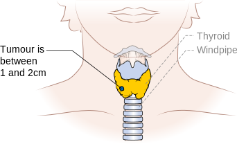
| T1b | Tumor >1 cm but ≤2 cm in greatest dimension, limited to the thyroid |
| T2 | Tumor >2 cm but ≤4 cm in greatest dimension, limited to the thyroid |
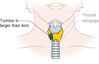
| T3 | Tumor >4 cm in greatest dimension limited to the thyroid or any tumor with minimal extrathyroid extension (e.g., extension to sternothyroid muscle or perithyroid soft tissues) |
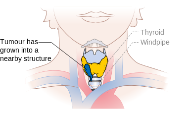
| T4a | Moderately advanced disease |
| Tumor of any size extending beyond the thyroid capsule to invade subcutaneous soft tissues, larynx, trachea, esophagus, or recurrent laryngeal nerve | |
| T4b | Very advanced disease |
| Tumor invades prevertebral fascia or encases carotid artery or mediastinal vessel |
| Stage | Description |
|---|---|
| NX | Regional lymph node cannot be assessed |
| N0 | No regional lymph node metastasis]] |
| N1 | Regional lymph node metastasis |
| N1a | Metastases to Level VI (pretracheal, paratracheal, and prelaryngeal/Delphian lymph nodes) |
| N1b | Metastases to unilateral, bilateral, or contralateral cervical (Levels I, II, III, IV, or V) or retropharyngeal or superior mediastinal lymph nodes (Level VII) |
| Stage | Description |
|---|---|
| M0 | No distant metastasis |
| M1 | Distant metastasis |
| Stage | T | N | M |
|---|---|---|---|
| Papillary thyroid carcinoma | |||
| YOUNGER THAN 45 YEARS | |||
| I | Any T | Any N | M0 |
| II | Any T | Any N | M1 |
| 45 YEARS AND OLDER | |||
| I | T1 | Any N | M1 |
| II | T2 | N0 | M0 |
| III | T3 | N0 | M0 |
| T1 | N1a | M0 | |
| T2 | N1a | M0 | |
| T3 | N1a | M0 | |
| IVA | T4a | N0 | M0 |
| T4a | N1a | M0 | |
| T1 | N1b | M0 | |
| T2 | N1b | M0 | |
| T3 | N1b | M0 | |
| T4a | N1b | M0 | |
| IVB | T4b | Any N | M0 |
| Stage IVC | Any T | Any N | M1 |
-
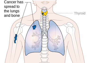
Stage M1 thyroid cancer
-
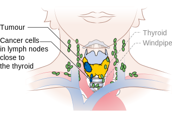
Stage N1a thyroid cancer
-
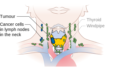
Stage N1b thyroid cancer
-
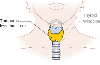
Stage T1a thyroid cancer
-
Stage T1b thyroid cancer
-
Stage T3 thyroid cancer
-
Stage T4a thyroid cancer
-
Stage T4b thyroid cancer


Key Biopsy Findings in Follicular Thyroid Cancer
- Characteristic Orphan Annie eye nuclear inclusions (nuclei with uniform staining, which appear empty)[3] and psammoma bodies on light microscopy. The former is useful in identifying the follicular variant of papillary thyroid carcinomas.[4]
- Histologically papillary carcinoma demonstrates 'delicate stalks of epithelial cells' which account for its name.
Reference
- ↑ Stage Information for Thyroid Cancer Cancer.gov (2015). http://www.cancer.gov/types/thyroid/hp/thyroid-treatment-pdq#link/stoc_h2_2- Accessed on October, 29 2015
- ↑ cancer.org > Thyroid Cancer By the American Cancer Society. In turn citing: AJCC Cancer Staging Manual (7th ed).
- ↑ "Papillary Carcinoma of Thyroid (Hi Pow)". University of Connecticut Health Center. Retrieved 2008-09-14.
- ↑ Yang GC, Liebeskind D, Messina AV (2001). "Ultrasound-guided fine-needle aspiration of the thyroid assessed by Ultrafast Papanicolaou stain: data from 1135 biopsies with a two- to six-year follow-up". Thyroid. 11 (6): 581–89. doi:10.1089/105072501750302895. PMID 11442006.