PEComa
| PEComa | |
 | |
|---|---|
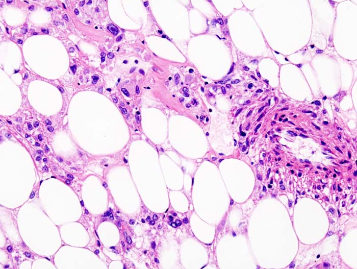
| Histopathologic image of renal angiomyolipoma. Nephrectomy specimen. H&E stain. | |
| eMedicine | orthoped/377 |
| MeSH | D054973 |
In oncology, PEComa, also PEC tumour and perivascular epithelioid cell tumour, is a type of mesenchymal tumour consisting of perivascular epithelioid cells (PECs).[1]
Histologic appearance
PECs consist of perivascular epithelioid cells with a clear/granular cytoplasm and central round nucleus without prominent nucleoli.
Immunohistochemical markers
PECs typically stain for melanocytic markers (HMB-45, Melan A (Mart 1), Mitf) and myogenic markers (actin).
PECs and other conditions
PECs bear significant histologic and immunohistochemical similarity to:
- angiomyolipoma,
- clear-cell sugar tumour (CCST),
- lymphangioleiomyomatosis, and,
- clear-cell myomelanocytic tumour of ligamentum teres/falciform ligament.
Thus, it has been advocated that the above could be classified PEComas.[1]
Etiology
The precursor cell of PEComas is currently unknown.[1] Genetically, PECs are linked to the tuberous sclerosis genes TSC1 and TSC2.