Giant cell tumor of bone overview
|
Giant cell tumor of bone Microchapters |
|
Differentiating Giant cell tumor of bone from other Diseases |
|---|
|
Diagnosis |
|
Treatment |
|
Case Studies |
|
Giant cell tumor of bone overview On the Web |
|
American Roentgen Ray Society Images of Giant cell tumor of bone overview |
|
Risk calculators and risk factors for Giant cell tumor of bone overview |
Editor-In-Chief: C. Michael Gibson, M.S., M.D. [1]
Overview
Giant cell tumor of the bone is a relatively uncommon tumor. It is characterized by the presence of multinucleated giant cells (osteoclast-like cells). These tumors are generally benign. In most patients, the tumors are slow to develop, but may recur locally in as many as 50% of cases. Metastasis to the lungs may occur.
Prevalence
Giant cell tumor of the bone accounts for 4-5% of primary bone tumors and 18.2% of benign bone tumors [1]. Giant cell tumors are mostly benign, however 5-10% of patients may have a malignant tumor.
Differential Diagnosis
A number of tumors have giant cells, but are not true benign giant cell tumors. These include
- Aneurysmal bone cyst
- Chondroblastoma
- Simple bone cyst
- Osteoid osteoma
- Osteoblastoma
- Osteosarcoma
- Giant cell reparative granuloma
- Brown tumor of hyperparathyroidism.
History and Symptoms
- Patients usually present with pain and limited range of motion caused by tumor's proximity to the joint space.
- There may be swelling as well, if the tumor has been growing for a long time.
- Some patients may be asymptomatic until they develop a pathologic fracture at the site of the tumor.
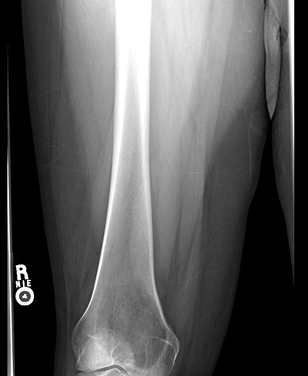
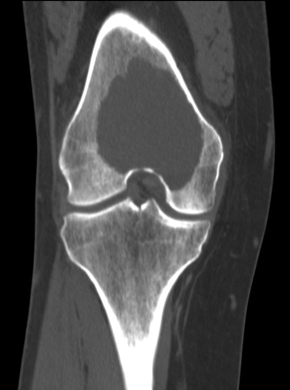
X-Ray
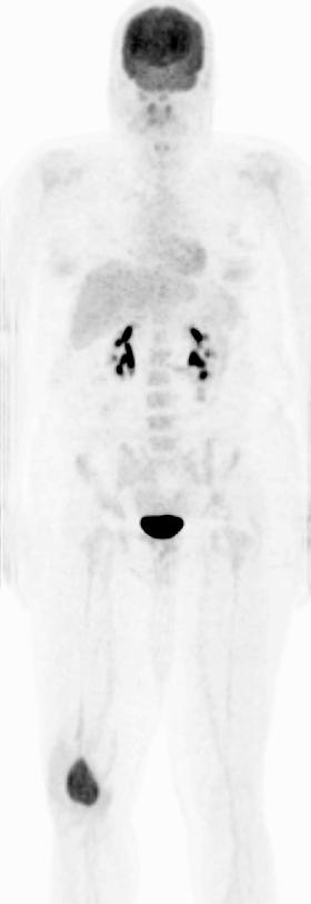
On x-ray, giant cell tumors (GCTs) have a metaepiphyseal location and grow to the articular surface of the involved bone [2]. They are distinguishable from other bony tumors in that GCTs usually have a non-sclerotic and sharply defined border. Because giant cell tumors are known to metastasize, when the diagnosis of giant cell tumor is suspected, a chest x-ray or CT may be needed.
(Images courtesy of RadsWiki)
-
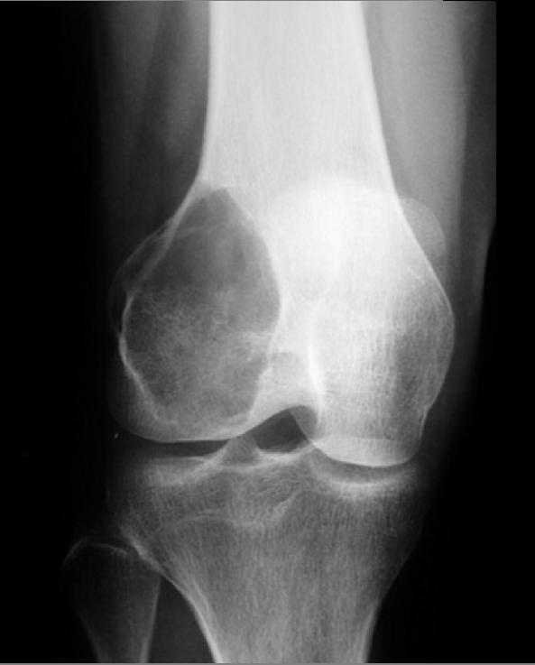
Giant cell tumor: Distal part of the femur
-
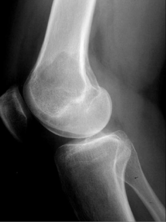
Giant cell tumor: Distal part of the femur


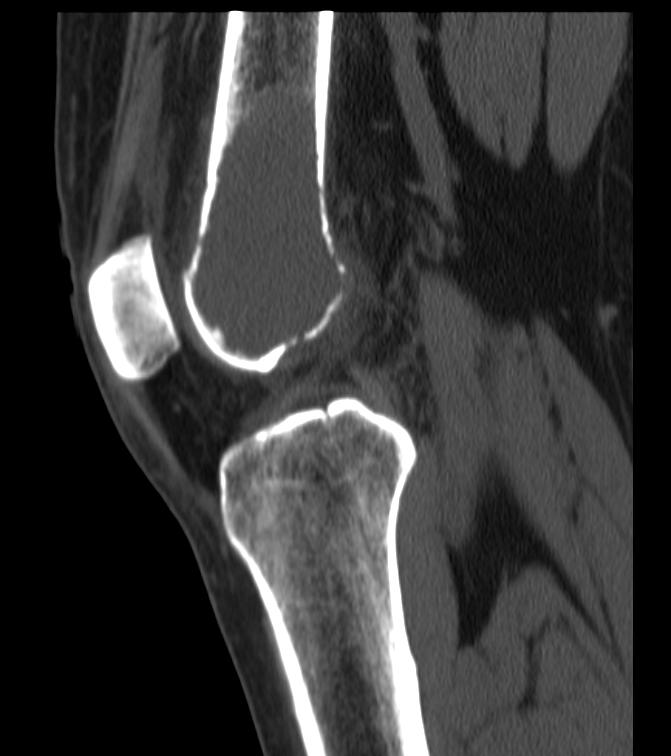
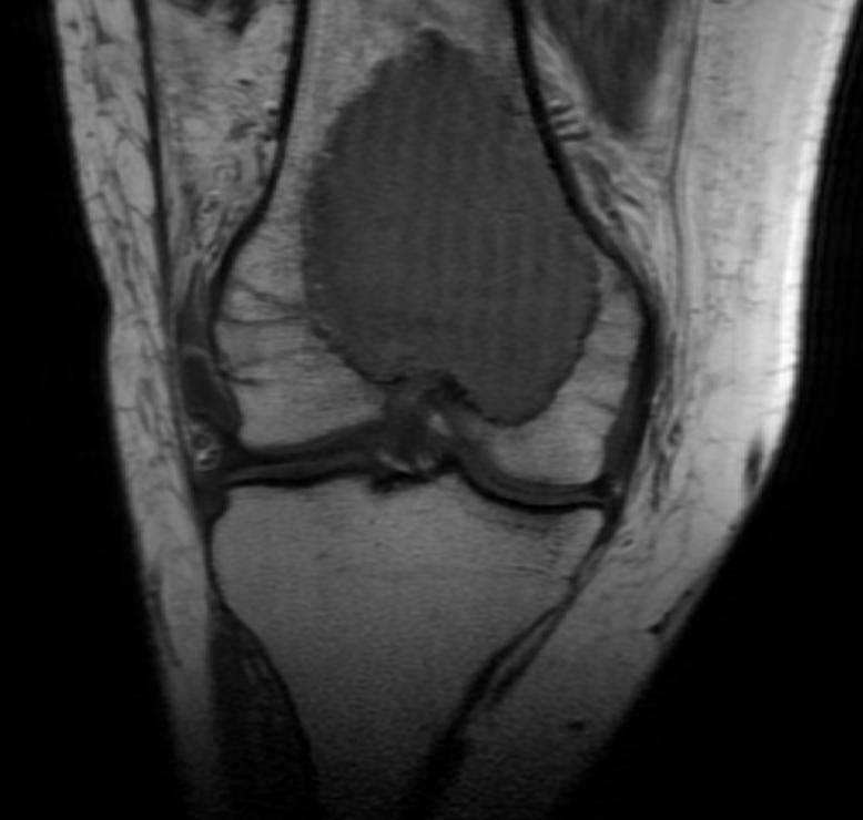
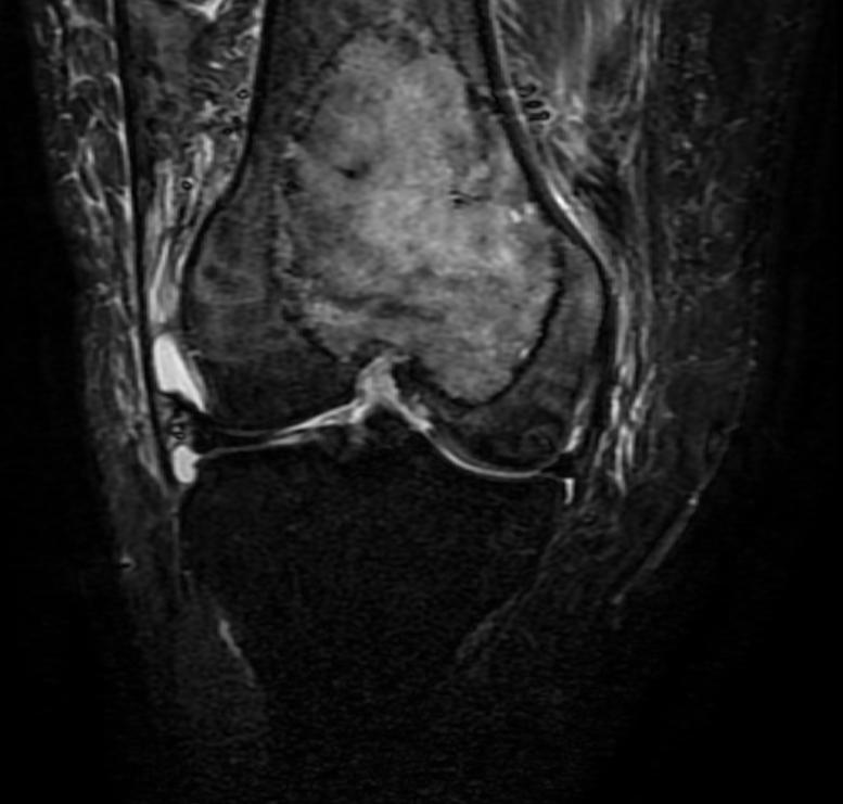
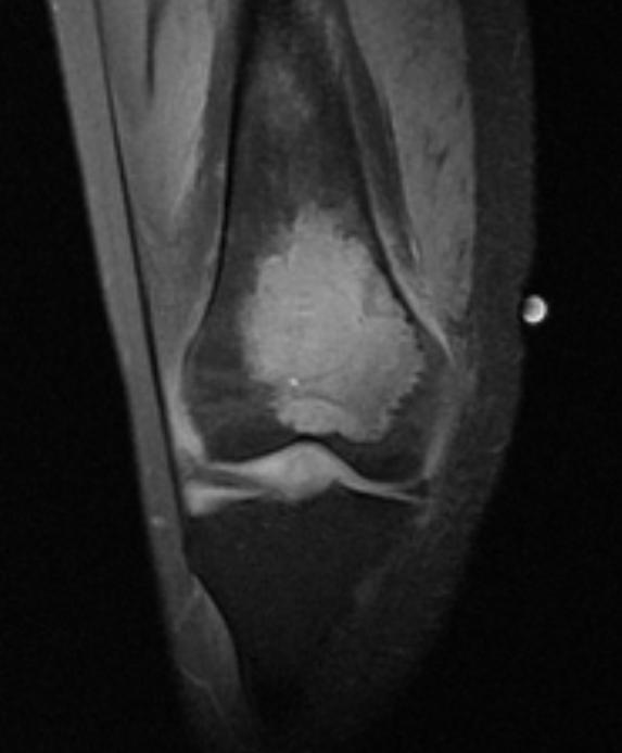
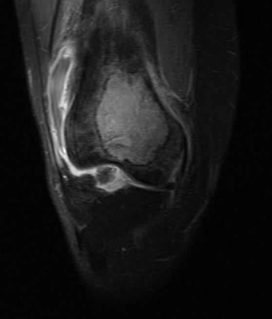
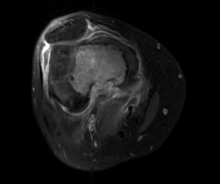
MRI
MRI can be used to assess intramedullary and soft tissue extension.
(Images courtesy of RadsWiki) Patient #1
-
Giant cell tumor: Distal part of the femur
-
Giant cell tumor: Distal part of the femur


-
Giant cell tumor: Distal part of the femur
-
Giant cell tumor: Distal part of the femur


-
Giant cell tumor: Distal part of the femur
-
Giant cell tumor: Distal part of the femur


-
Giant cell tumor: Distal part of the femur
-
Giant cell tumor: Distal part of the femur
-
Giant cell tumor: Distal part of the femur


Radiation Therapy
Patients with tumors that are not amenable to surgery are treated with radiation therapy [3].
References
- ↑ Gamberi G, Serra M, Ragazzini P, Magagnoli G, Pazzaglia L, Ponticelli F, Ferrari C, Zanasi M, Bertoni F, Picci P, Benassi MS (2003). "Identification of markers of possible prognostic value in 57 giant cell tumors of bone". Oncology Reports. 10 (2): 351–6. PMID 12579271. Retrieved 2012-01-18.
- ↑ Murphey MD, Nomikos GC, Flemming DJ, Gannon FH, Temple HT, Kransdorf MJ (2001). "From the archives of AFIP. Imaging of giant cell tumor and giant cell reparative granuloma of bone: radiologic-pathologic correlation". Radiographics : a Review Publication of the Radiological Society of North America, Inc. 21 (5): 1283–309. PMID 11553835. Retrieved 2012-01-18.
- ↑ Mendenhall WM, Zlotecki RA, Scarborough MT, Gibbs CP, Mendenhall NP (2006). "Giant cell tumor of bone". American Journal of Clinical Oncology. 29 (1): 96–9. doi:10.1097/01.coc.0000195089.11620.b7. PMID 16462511. Retrieved 2012-01-18. Unknown parameter
|month=ignored (help)