Renal cell carcinoma pathophysiology
Editor-In-Chief: C. Michael Gibson, M.S., M.D. [1]
|
Renal cell carcinoma Microchapters |
|
Diagnosis |
|---|
|
Treatment |
|
Case Studies |
|
Renal cell carcinoma pathophysiology On the Web |
|
American Roentgen Ray Society Images of Renal cell carcinoma pathophysiology |
|
Risk calculators and risk factors for Renal cell carcinoma pathophysiology |
Overview
Renal cell carcinoma is the most common form of kidney cancer arising from the renal tubule. It is the most common type of kidney cancer in adults. Initial treatment is surgery. It is notoriously resistant to radiation therapy and chemotherapy, although some cases respond to immunotherapy. The advent of targeted cancer therapies such as sunitinib has vastly improved the outlook for treatment of RCC.
Pathophysiology
Genetics
Recent genetic studies have altered the approaches of understanding renal cell carcinoma. [1][2][3]
- VHL and others on chromosome 3 - Clear cell carcinoma
- MET, PRCC - Papillary carcinoma
- Other associated genes include TRC8, OGG1, HNF1A, HNF1B, TFE3, RCCP3, and RCC17.
Gross Pathology
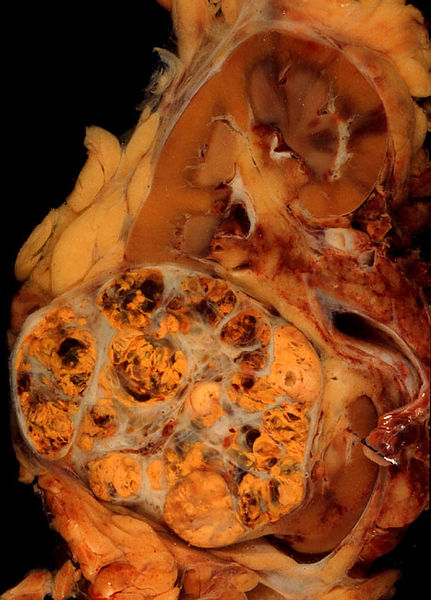
Gross examination shows a hypervascular lesion in the renal cortex, which is frequently multilobulated, yellow (because of the lipid accumulation) and calcified.

Light Microscopy
Light microscopy shows tumor cells forming cords, papillae, tubules or nests, and are atypical, polygonal and large. Because these cells accumulate glycogen and lipids, their cytoplasm appear "clear", lipid-laden, the nuclei remain in the middle of the cells, and the cellular membrane is evident. Some cells may be smaller, with eosinophilic cytoplasm, resembling normal tubular cells. The stroma is reduced, but well vascularized. The tumor grows in large front, compressing the surrounding parenchyma, producing a pseudocapsule.[4]
Secretion of vasoactive substances (e.g. renin) may cause arterial hypertension, and release of erythropoietin may cause polycythemia (increased production of red blood cells).
References
- ↑ Reuter VE, Presti JC (2000). "Contemporary approach to the classification of renal epithelial tumors". Semin. Oncol. 27 (2): 124–37. PMID 10768592. Unknown parameter
|month=ignored (help) - ↑ Bodmer D, van den Hurk W, van Groningen JJ; et al. (2002). "Understanding familial and non-familial renal cell cancer". Hum. Mol. Genet. 11 (20): 2489–98. PMID 12351585. Unknown parameter
|month=ignored (help) - ↑ Cotran, Ramzi S.; Kumar, Vinay; Fausto, Nelson; Nelso Fausto; Robbins, Stanley L.; Abbas, Abul K. (2005). Robbins and Cotran pathologic basis of disease. St. Louis, Mo: Elsevier Saunders. p. 1016. ISBN 0-7216-0187-1.
- ↑ http://www.pathologyatlas.ro/Renal%20Clear%20Cell%20Carcinoma.html