Gangrene classification: Difference between revisions
| Line 18: | Line 18: | ||
The early signs of dry gangrene are a dull ache and sensation of coldness in the affected area along with [[pallor]] of the flesh. If caught early, the process can sometimes be reversed by vascular surgery. However, if necrosis sets in, the affected tissue must be removed just as with wet gangrene. | The early signs of dry gangrene are a dull ache and sensation of coldness in the affected area along with [[pallor]] of the flesh. If caught early, the process can sometimes be reversed by vascular surgery. However, if necrosis sets in, the affected tissue must be removed just as with wet gangrene. | ||
<div align=" | <div align="center"> | ||
<gallery heights="175" widths="175"> | <gallery heights="175" widths="175"> | ||
Image:ULCERCELLULITIS1.JPG|Diabetic [[ulcer]]ation with central "dry" gangrene and toward the edges wet gangrene with some ascending [[cellulitis]] <br> (Image courtesy of Charlie Goldberg, M.D., UCSD School of Medicine and VA Medical Center, San Diego, CA) | Image:ULCERCELLULITIS1.JPG|Diabetic [[ulcer]]ation with central "dry" gangrene and toward the edges wet gangrene with some ascending [[cellulitis]] <br> (Image courtesy of Charlie Goldberg, M.D., UCSD School of Medicine and VA Medical Center, San Diego, CA) | ||
Revision as of 20:31, 27 February 2013
|
Gangrene Microchapters |
|
Diagnosis |
|---|
|
Treatment |
|
Case Studies |
|
Gangrene classification On the Web |
|
American Roentgen Ray Society Images of Gangrene classification |
|
Risk calculators and risk factors for Gangrene classification |
Editor-In-Chief: C. Michael Gibson, M.S., M.D. [1]
References
Overview
Three main types of gangrene in clude wet, dry and gas gangrene. Sometimes it can be classified according to its site.
Types of gangrene
Dry gangrene
Dry gangrene begins at the distal part of the limb due to ischemia and often occurs in the toes and feet of elderly patients due to arteriosclerosis. Dry gangrene spreads slowly until it reaches the point where the blood supply is inadequate to keep tissue viable. Macroscopically, the affected part is dry, shrunken and dark black, resembling mummified flesh. The dark coloration is due to liberation of hemoglobin from hemolyzed red blood cells which is acted upon by hydrogen sulfide (H2S) produced by the bacteria, resulting in formation of black iron sulfide that remains in the tissues[1]. The line of separation usually brings about complete separation with eventual falling off of the gangrenous tissue if it is not removed surgically.
If the blood flow is interrupted for a reason other than severe bacterial infection, the result is a case of dry gangrene. People with impaired peripheral blood flow, such as diabetics, are at greater risk of contracting dry gangrene.
The early signs of dry gangrene are a dull ache and sensation of coldness in the affected area along with pallor of the flesh. If caught early, the process can sometimes be reversed by vascular surgery. However, if necrosis sets in, the affected tissue must be removed just as with wet gangrene.
-
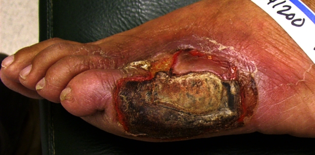
Diabetic ulceration with central "dry" gangrene and toward the edges wet gangrene with some ascending cellulitis
(Image courtesy of Charlie Goldberg, M.D., UCSD School of Medicine and VA Medical Center, San Diego, CA)

Wet gangrene
Wet gangrene occurs in naturally moist tissue and organs such as the mouth, bowel, lungs, cervix, and vulva. Bedsores occurring on body parts such as the sacrum, buttocks and heels—although not necessarily moist areas—are also categorized as wet gangrene infections. In wet gangrene, the tissue is infected by saprogenic microorganisms (Bac.perfringes, fusiformis, putrificans, etc.), which cause tissue to swell and emit a fetid smell. Wet gangrene usually develops rapidly due to blockage of venous and/or arterial blood flow. The affected part is saturated with stagnant blood which promotes the rapid growth of bacteria. The toxic products formed by bacteria are absorbed causing systemic manifestation of septicemia and finally death. Macroscopically, the affected part is edematous, soft, putrid, rotten and dark. The darkness in wet gangrene occurs due to the same mechanism as in dry gangrene.
-
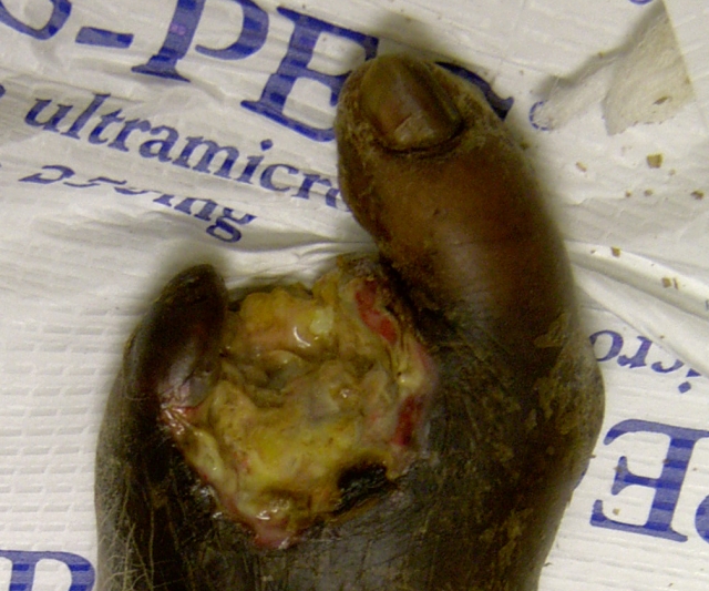
Diabetic with severe infection and loss of toes - wet gangrene in center.
(Image courtesy of Charlie Goldberg, M.D., UCSD School of Medicine and VA Medical Center, San Diego, CA)

Gas gangrene
Gas gangrene is a bacterial infection that produces gas within tissues. It is a deadly form of gangrene usually caused by Clostridium perfringens bacteria. Infection spreads rapidly as the gases produced by bacteria expand and infiltrate healthy tissue in the vicinity. Because of its ability to quickly spread to surrounding tissues, gas gangrene should be treated as a medical emergency.
Gas gangrene is caused by a bacterial exotoxin-producing clostridial species, which are mostly found in soil and other anaerobes (e.g. Bacteroides and anaerobic streptococci). These environmental bacteria may enter the muscle through a wound and subsequently proliferate in necrotic tissue and secrete powerful toxins. These toxins destroy nearby tissue, generating gas at the same time. A gas composition of 5.9% hydrogen, 3.4% carbon dioxide, 74.5% nitrogen and 16.1% oxygen was reported in one clinical case.[2]
Gas gangrene can cause necrosis, gas production, and sepsis. Progression to toxemia and shock is often very rapid.
Specific gangrenes
- Noma is a gangrene of the face.
- Necrotizing fasciitis affects the deeper layers of the skin.
- Fournier gangrene usually affects the male genitals.