Vestibular nuclei
Template:Infobox Brain Editor-In-Chief: C. Michael Gibson, M.S., M.D. [1]
Overview
The vestibular nuclei are the cranial nuclei for the vestibular nerve.
Subnuclei
There are 4 subnuclei; they are situated at the floor of the fourth ventricle.
- The medial vestibular nucleus (dorsal or chief vestibular nucleus), corresponding to the lower part of the area acustica in the rhomboid fossa; the caudal end of this nucleus is sometimes termed the descending or spinal vestibular nucleus.
- The lateral vestibular nucleus or nucleus of Deiters, consisting of large cells and situated in the lateral angle of the rhomboid fossa; the dorso-lateral part of this nucleus is sometimes termed the nucleus of Bechterew.
- Modern sources also include a inferior vestibular nucleus and superior vestibular nucleus
Path from medial and lateral nuclei
The fibers of the vestibular nerve enter the medulla oblongata on the medial side of those of the cochlear, and pass between the inferior peduncle and the spinal tract of the trigeminal.
They then divide into ascending and descending fibers. The latter end by arborizing around the cells of the medial nucleus, which is situated in the area acustica of the rhomboid fossa. The ascending fibers either end in the same manner or in the lateral nucleus, which is situated lateral to the area acustica and farther from the ventricular floor.
Some of the axons of the cells of the lateral nucleus, and possibly also of the medial nucleus, are continued upward through the inferior peduncle to the roof nuclei of the opposite side of the cerebellum, to which also other fibers of the vestibular root are prolonged without interruption in the nuclei of the medulla oblongata.
A second set of fibres from the medial and lateral nuclei end partly in the tegmentum, while the remainder ascend in the medial longitudinal fasciculus to arborize around the cells of the nuclei of the oculomotor nerve.
Fibres from the lateral vestibular nucleus also pass via the vestibulospinal tract, to anterior horn cells at many levels in the spinal cord, in order to co-ordinate head and trunk movements.
See also
Additional images
-
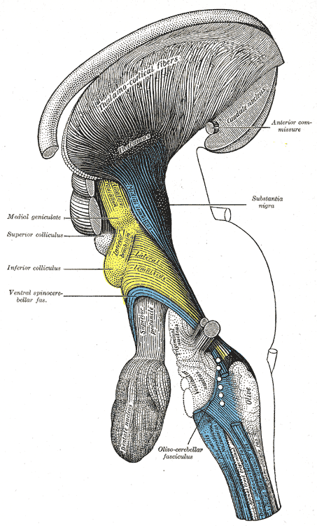 Deep dissection of brain-stem. Lateral view.
 -
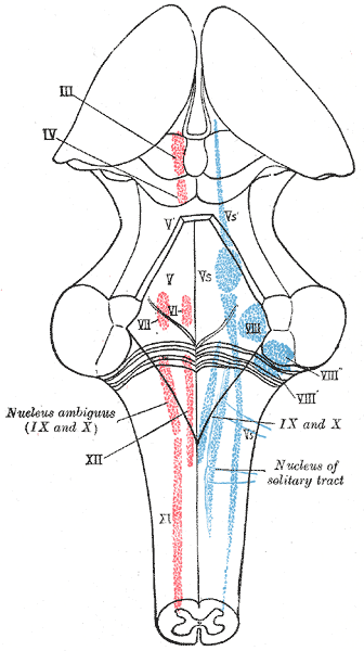 The cranial nerve nuclei schematically represented; dorsal view. Motor nuclei in red; sensory in blue.
 -
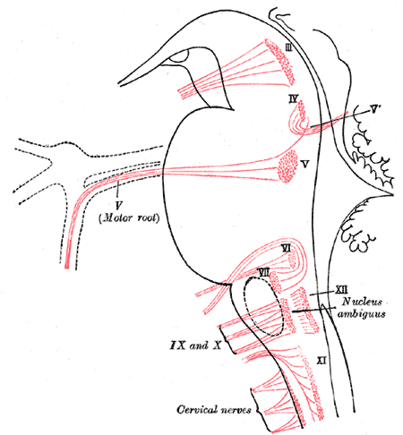 Nuclei of origin of cranial motor nerves schematically represented; lateral view.
 -
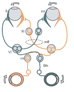
 Primary terminal nuclei of the afferent (sensory) cranial nerves schematically represented; lateral view.
 -


External links
Template:Gray's Template:Rhombencephalon Template:WH Template:WS