Umbilical artery
|
WikiDoc Resources for Umbilical artery |
|
Articles |
|---|
|
Most recent articles on Umbilical artery Most cited articles on Umbilical artery |
|
Media |
|
Powerpoint slides on Umbilical artery |
|
Evidence Based Medicine |
|
Clinical Trials |
|
Ongoing Trials on Umbilical artery at Clinical Trials.gov Trial results on Umbilical artery Clinical Trials on Umbilical artery at Google
|
|
Guidelines / Policies / Govt |
|
US National Guidelines Clearinghouse on Umbilical artery NICE Guidance on Umbilical artery
|
|
Books |
|
News |
|
Commentary |
|
Definitions |
|
Patient Resources / Community |
|
Patient resources on Umbilical artery Discussion groups on Umbilical artery Patient Handouts on Umbilical artery Directions to Hospitals Treating Umbilical artery Risk calculators and risk factors for Umbilical artery
|
|
Healthcare Provider Resources |
|
Causes & Risk Factors for Umbilical artery |
|
Continuing Medical Education (CME) |
|
International |
|
|
|
Business |
|
Experimental / Informatics |
Editor-In-Chief: C. Michael Gibson, M.S., M.D. [1]
Associate Editor-In-Chief: Cafer Zorkun, M.D., Ph.D. [2]
Overview
The umbilical artery is a paired artery (with one for each half of the body) that is found in the abdominal and pelvic regions. In the fetus, it extends into the umbilical cord.
Umbilical arteries in the fetus
Umbilical arteries carry deoxygenated blood from the fetus to the placenta in the umbilical cord. There are usually two umbilical arteries present together with one umbilical vein in the cord. The umbilical arteries are actually the latter of the internal iliac arteries that supply the hind limbs with blood in the fetus. The umbilical arteries surround the urinary bladder and then carry all the deoxygenated blood out of the fetus through the umbilical cord.
The umbilical artery is the only artery present in the human body, aside from the pulmonary arteries, which carries deoxygenated blood.
Single Umbilical Artery
Occasionally, there is only the one single umbilical artery (SUA) present in the cord. This abnormality is typically more common in multiple births, but can appear with singletons as well. Babies with SUA may have a higher likelihood of having other congenital abnormalities. However, additional testing (high level ultrasound scans) can rule out these abnormalities prior to birth and alleviate parental anxiety. Genetic counseling may be useful, too, especially when weighing the pros and cons of more invasive procedures such as chorionic villus sampling and amniocentesis.
It is important to note that a diagnosis of SUA, while anxiety provoking, is in no way a guarantee that a fetus is at risk for other problems in utero or after birth. Especially encouraging are cases in which no other soft markers for congenital abnormalities are visible via ultrasound. In fact, medical literature suggests that 1 in 100 newborns have it, making it the most common umbilical abnormality. Prior to ultrasound technology, the only method for determining the presence of such a cord was at birth, following an examination of the placenta. Given that the vast majority of expectant mothers do not receive the kind of advanced ultrasound scanning required to confirm SUA in utero, most cases may never detected antenatally even today.
In the United States, doctors and midwives typically encourage parents to take the added precaution of having regular growth scans near term to rule out intrauterine growth restriction, which can happen on occasion and warrant intervention. Yet the majority of growth restricted infants with the abnormality also have other defects. Echocardiograms may be advised late in the pregnancy to ensure the heart is functioning properly. Finally, neonates with the finding may also have a higher occurrence of kidney problems, so close examination of the infant may be warranted shortly after birth. Among SUA infants, there is a slightly elevated risk for post-natal urinary infections.
Umbilical artery in the adult
The umbilical artery is a branch of the anterior division of the internal iliac artery and represents the patent (open) part of the embryonic umbilical artery. (The non-patent obliterated part of the artery is the medial umbilical ligament.) The umbilical artery is found in the pelvis, and gives rise to the superior vesical arteries. In males, it also gives rise to the artery to the ductus deferens.
Additional images
-
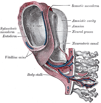 Model of human embryo 1.3 mm. long.
 -
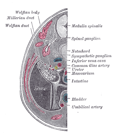 Transverse section of human embryo eight and a half to nine weeks old.
 -
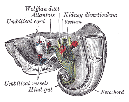 Tail end of human embryo twenty-five to twenty-nine days old.
 -
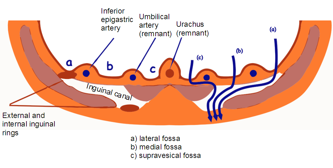 Inguinal fossae


External links
- Template:GPnotebook
- Template:SUNYAnatomyLabs - "The Female Pelvis: Branches of Internal Iliac Artery"
Template:Arteries of thorax and abdomen Template:Embryology Template:Development of circulatory system