Scalp
Template:Infobox Anatomy Editor-In-Chief: C. Michael Gibson, M.S., M.D. [1]
Overview
The scalp is the anatomical area bordered by the face anteriorly and the neck to the sides and posteriorly.
Layers
It is usually described as having five layers, which can be remembered with the mnemonic "SCALP":[1]
- S: The skin on the head from which head hair grows. It is richly supplied with blood vessels and can be subject to such conditions as dandruff and cutis verticis gyrata.
- C: Connective tissue. a thin layer of fat and fibrous tissue lies beneath the skin
- A: The aponeurosis (or galea aponeurotica) is the next layer. It is a tough layer of dense fibrous tissue which runs from the frontalis muscle anteriorly to the occipitalis posteriorly
- L: The loose areolar connective tissue layer provides an easy plane of separation between the upper three layers and the pericranium. In scalping the scalp is torn off through this layer. It also provides a plane of access in craniofacial surgery and neurosurgery. This layer is sometimes referred to as the "Danger Zone" because of the ease by which infectious agents can spread through it to emissary veins which then drain into the cranium. The loose areolar tissue in this layer is made up of random collagen I bundles, collagen III and is highly vascular and cellular. It will also be rich in glycosaminoglycans (GAGs) and will be constituted of more matrix than fibers.
- P: The pericranium is the periosteum of the skull bones and provides nutrition to the bone and the capacity for repair. It may be lifted from the bone to allow removal of bone windows (craniotomy).
Blood supply
The blood supply of the scalp is via four pairs of arteries, two from the external carotid and two from the internal carotid:
- internal carotid
- the supratrochlear artery to the midline forehead
- the supraorbital artery to the lateral forehead and scalp as far up as the vertex
- external carotid
- the superficial temporal artery which gives frontal and parietal branches to supply much of the scalp
- the occipital artery which runs from posteriorly to supply much of the back of the scalp.
Innervation
The scalp is innervated by the following:[2]
- Supratrochlear nerve and the supraorbital nerve from the ophthalmic division of the trigeminal nerve
- Greater occipital nerve (C2) posteriorly up to the vertex
- Lesser occipital nerve (C3) behind the ear.
- Zygomaticotemporal nerve from the maxillary division of the trigeminal nerve supplying the hairless temple
- Auriculotemporal nerve from the mandibular division of the trigeminal nerve
Role in aesthetics
The scalp plays an important role in the aesthetics of the face. Androgenic alopecia, or male pattern hair loss, is a common cause of concern to men. It may be treated by medication (eg finasteride) or hair transplantation with variable success. If the scalp is heavy and loose, a common change with aging, the forehead may be low, heave and deeply lined. The brow lift procedure aims to address these concerns.
Pathology
The scalp is a common site for the development of tumours including:
- epidermoid cyst
- pilar cyst
- actinic keratosis and squamous cell carcinoma
- basal cell carcinoma
- merkel cell tumours
See also
- Trichology -- the scientific study of hair and scalp
- Trichodynia -- burning scalp syndrome
Additional images
-
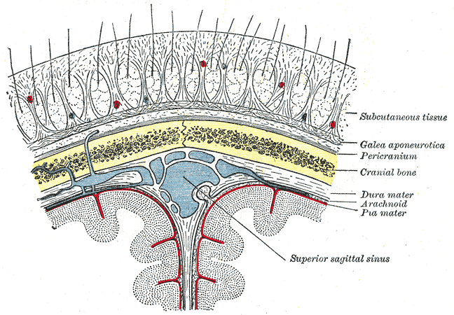
Diagrammatic section of scalp.

References
Template:Head and neck general
External links
- Histology image: 08601ooa – Histology Learning System at Boston University - "Integument: scalp, transverse"
- Histology image: 08801ooa – Histology Learning System at Boston University - "Integument: scalp"
- Template:NormanAnatomy
de:Kopfschwarte eo:Skalpo it:cuoio capelluto ko:머리가죽 nl:Kruin fi:Päänahka sv:Skalp