Pseudoaneurysm
|
WikiDoc Resources for Pseudoaneurysm |
|
Articles |
|---|
|
Most recent articles on Pseudoaneurysm Most cited articles on Pseudoaneurysm |
|
Media |
|
Powerpoint slides on Pseudoaneurysm |
|
Evidence Based Medicine |
|
Clinical Trials |
|
Ongoing Trials on Pseudoaneurysm at Clinical Trials.gov Trial results on Pseudoaneurysm Clinical Trials on Pseudoaneurysm at Google
|
|
Guidelines / Policies / Govt |
|
US National Guidelines Clearinghouse on Pseudoaneurysm NICE Guidance on Pseudoaneurysm
|
|
Books |
|
News |
|
Commentary |
|
Definitions |
|
Patient Resources / Community |
|
Patient resources on Pseudoaneurysm Discussion groups on Pseudoaneurysm Patient Handouts on Pseudoaneurysm Directions to Hospitals Treating Pseudoaneurysm Risk calculators and risk factors for Pseudoaneurysm
|
|
Healthcare Provider Resources |
|
Causes & Risk Factors for Pseudoaneurysm |
|
Continuing Medical Education (CME) |
|
International |
|
|
|
Business |
|
Experimental / Informatics |
Editor-In-Chief: C. Michael Gibson, M.S., M.D. [1]
Overview
A pseudoaneurysm, also known as a false aneurysm, is an outpouching of a blood vessel involving a defect in the two innermost tissue layers (tunica intima and media). The outermost layer (adventia) may be intact, or alternatively, all three layers may be damaged, with bleeding contained by a blood clot or surrounding structures. True aneurysms in contrast, are vascular outpouchings containing all three tissue layers.
Epidemiology and Demographics
Given the increase in invasive cardiac procedures, damage to all three layers is the more common source of pseudoaneurysm in modern medical settings. Damage to the two innermost layers is more commonly seen following trauma to a vessel. Femoral pseudoaneurysms may complicate up to 8% of vascular interventional procedures.
Pseudoaneuryms of Structures other than Blood Vessels: Left Ventricular Pseudoaneurysms
A pseudoaneurysm may also occur in a chamber of the heart following myocardial damage due to ischemia or trauma. An pseudoaneurysm of the left ventricle is a potentially lethal complication from a heart attack. After a heart attack, the left ventricular wall of the heart, may rupture.
Although aneurysms and left ventricular aneurysms may involve any wall segment, aneurysms in the posterolateral wall are frequently due to pseudoaneurysms. In contrast, the most common location for a true left ventricular aneurysm involves the apex of the heart.
Diagnosis
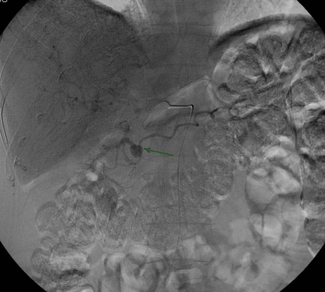
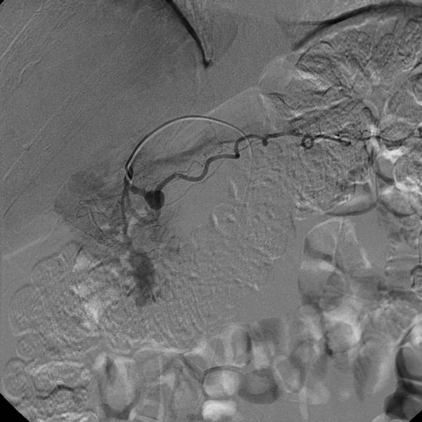
Patient #1: Gastroduodenal artery pseudoaneurysm
-

-
-
Angiogram
-
-
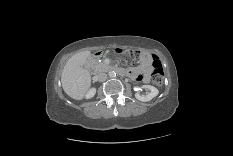 Post Embolization CT


Ultrasound
- Yin-yang sign: Swirling blood flow pattern within a cystic structure.
- To-and-fro flow: to represents blood entering the pseudoaneurysm in systole and fro represents blood exiting the pseudoaneurysm during diastole.
Treatment
Small pseudoaneurysms can spontaneously clot, while others need definitive treatment. Surgery is considered the gold-standard treatment, although is not without risk in patients with severe cardiovascular disease. Less invasive treatment options, such as Duplex ultrasound-guided compression and percutaneous thrombin injection are available, however, evidence of their efficacy is somewhat limited.
Prevention
External compression, either manual or device usually do not prevent the pseudoaneurysm from forming once the tunica intima has been damaged.