Mitral stenosis electrocardiogram
Editor-In-Chief: C. Michael Gibson, M.S., M.D. [1]; Associate Editor-In-Chief: Cafer Zorkun, M.D., Ph.D. [2]; Varun Kumar, M.B.B.S.; Lakshmi Gopalakrishnan, M.B.B.S.
|
Mitral Stenosis Microchapters |
|
Diagnosis |
|---|
|
Treatment |
|
Case Studies |
|
Mitral stenosis electrocardiogram On the Web |
|
American Roentgen Ray Society Images of Mitral stenosis electrocardiogram |
|
Risk calculators and risk factors for Mitral stenosis electrocardiogram |
Overview
The electrocardiogram (ECG) in mitral stenosis might have no significant abnormalities. Findings suggestive of left atrial enlargement and hypertrophy might be present, such as a broad, bifid P wave in lead II (referred to as P mitrale) and an enlarged terminal negative portion of the P wave in V1. The ECG might demonstrate findings of pulmonary hypertension and right ventricular hypertophy. Atrial fibrillation is not an uncommon finding among patients with mitral stenosis.
Electrocardiogram
Left Atrial Enlargement
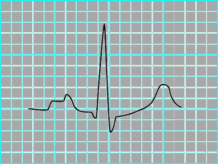
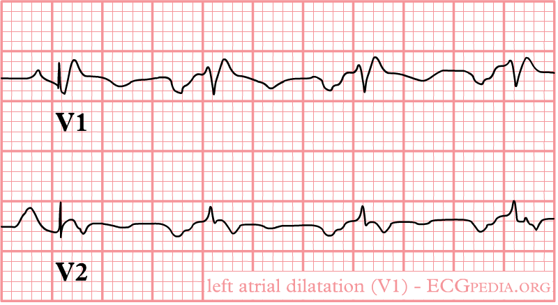
Left atrial enlargement produces a broad, bifid P wave in lead II (P mitrale) and enlarges the terminal negative portion of the P wave in V1.[1][2]
Shown below is an ECG depicting the following in lead II:[3]

Shown below is an ECG depicting the following in lead V1:[4]
- Biphasic P wave with terminal negative portion > 40 ms duration
- Biphasic P wave with terminal negative portion > 1mm deep

Copyleft image obtained courtesy of, http://en.ecgpedia.org/wiki/Main_Page
Right Ventricular Hypertrophy
ECG findings suggestive of right ventricular hypertrophy include:
- Right axis deviation of +90 degrees or more
- RV1 = 7 mm or more
- RV1 + SV5 or SV6 = 10 mm or more
- R/S ratio in V1 = 1.0 or more
- S/R ratio in V6 = 1.0 or more
- Late intrinsicoid deflection in V1 (0.035+)
- Incomplete RBBB pattern
- ST T strain pattern in 2,3,aVF
- P pulmonale or Right atrial enlargement or P congenitale
- S1 S2 S3 pattern in children
- Tall R wave in V1 or qR in V1
- R wave greater than S wave in V1
- R wave progression reversal
- Inverted T wave in the anterior precordial leads
Rigth Axis Deviation
In right axis deviation, the mean QRS axis in the frontal plane moves toward the right as pulmonary hypertension worsens.
Atrial Fibrillation
Atrial fibrillation is commonly seen with mitral stenosis. Atrial fibrillation is characterized by an irregularly irregular rhythm with absent P waves.
EKG Examples of Mitral stenosis
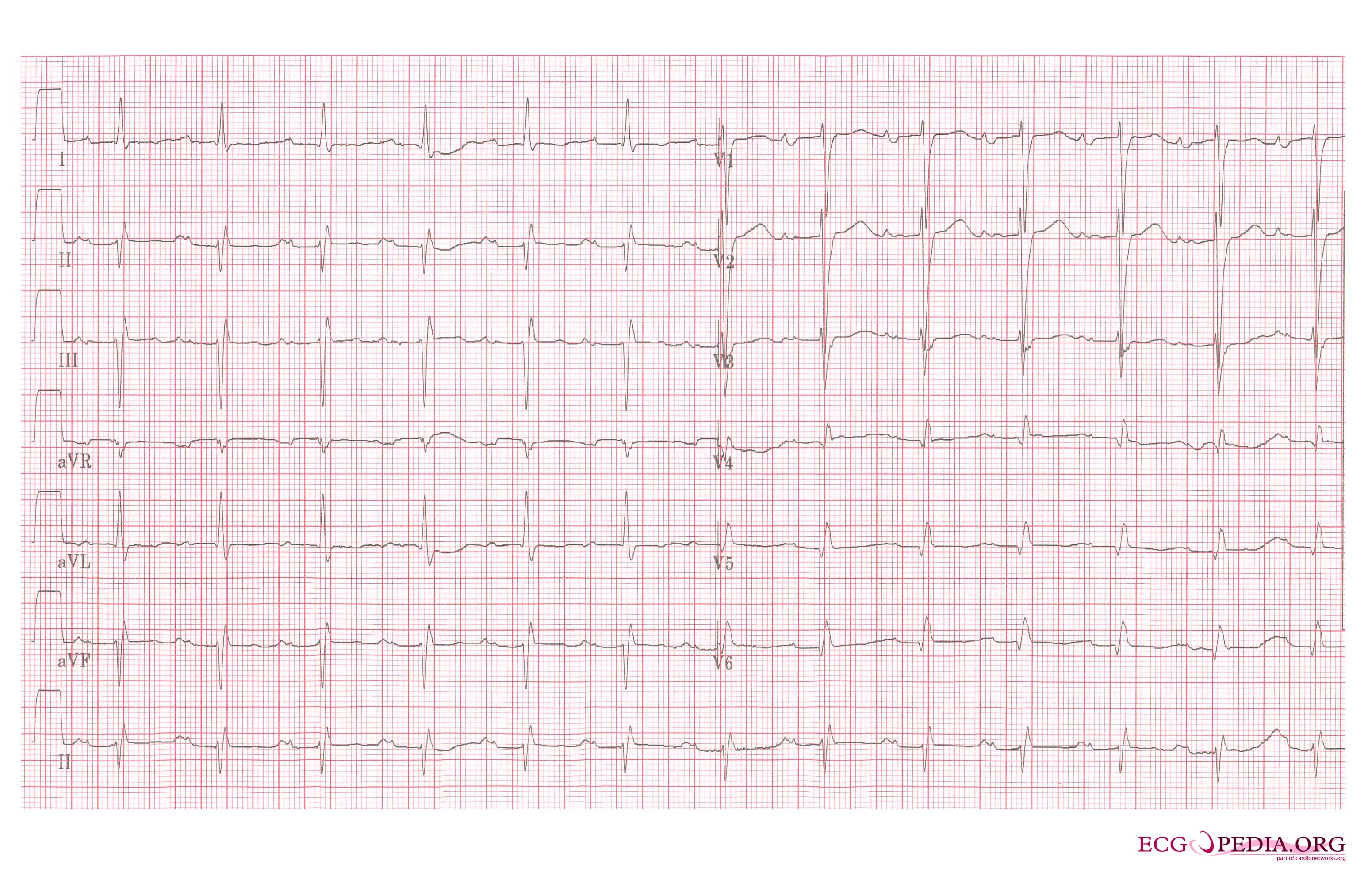
Shown below is an ECG image of a patient with mitral stenosis.

Copyleft image obtained courtesy of, http://en.ecgpedia.org/wiki/Main_Page
References
- ↑ Maganti K, Rigolin VH, Sarano ME, Bonow RO (2010). "Valvular heart disease: diagnosis and management". Mayo Clin Proc. 85 (5): 483–500. doi:10.4065/mcp.2009.0706. PMC 2861980. PMID 20435842.
- ↑ TROUNCE JR (1952). "The electrocardiogram in mitral stenosis". Br Heart J. 14 (2): 185–92. PMC 479442. PMID 14916061.
- ↑ Pérez-Riera, Andrés Ricardo; de Abreu, Luiz Carlos; Barbosa-Barros, Raimundo; Grindler, José; Fernandes-Cardoso, Acácio; Baranchuk, Adrian (2016). "P-wave dispersion: an update". Indian Pacing and Electrophysiology Journal. 16 (4): 126–133. doi:10.1016/j.ipej.2016.10.002. ISSN 0972-6292.
- ↑ Pérez-Riera AR, de Abreu LC, Barbosa-Barros R, Grindler J, Fernandes-Cardoso A, Baranchuk A (2016). "P-wave dispersion: an update". Indian Pacing Electrophysiol J. 16 (4): 126–133. doi:10.1016/j.ipej.2016.10.002. PMC 5197451. PMID 27924760.