Lysosome


|
WikiDoc Resources for Lysosome |
|
Articles |
|---|
|
Most recent articles on Lysosome |
|
Media |
|
Evidence Based Medicine |
|
Clinical Trials |
|
Ongoing Trials on Lysosome at Clinical Trials.gov Clinical Trials on Lysosome at Google
|
|
Guidelines / Policies / Govt |
|
US National Guidelines Clearinghouse on Lysosome
|
|
Books |
|
News |
|
Commentary |
|
Definitions |
|
Patient Resources / Community |
|
Directions to Hospitals Treating Lysosome Risk calculators and risk factors for Lysosome
|
|
Healthcare Provider Resources |
|
Causes & Risk Factors for Lysosome |
|
Continuing Medical Education (CME) |
|
International |
|
|
|
Business |
|
Experimental / Informatics |
Editor-In-Chief: C. Michael Gibson, M.S., M.D. [1]
Associate Editor-In-Chief: Cafer Zorkun, M.D., Ph.D. [2]
Overview
Lysosomes are organelles that contain digestive enzymes (acid hydrolases). They digest excess or worn out organelles, food particles, and engulfed viruses or bacteria. The membrane surrounding a lysosome prevents the digestive enzymes inside from destroying the cell. Lysosomes fuse with vacuoles and dispense their enzymes into the vacuoles, digesting their contents. They are built in the Golgi apparatus. The name lysosome derives from the Greek words lysis, which means dissolution or destruction, and soma, which means body. They are frequently nicknamed "suicide-bags" or "suicide-sacs" by cell biologists due to their role in autolysis. Lysosomes were discovered by the Belgian cytologist Christian de Duve in 1949.
Acidic environment
At pH 4.8, the interior of the lysosomes is more acidic than the cytosol (pH 7.2). The lysosome single membrane stabilizes the low pH by pumping in protons (H+) from the cytosol via proton pumps and chloride ion channels. The membrane also protects the cytosol, and therefore the rest of the cell, from the degradative enzymes within the lysosome. For this reason, should a lysosome's acid hydrolases leak into the cytosol, their potential to damage the cell will be reduced, because they will not be at their optimum pH!
Enzymes
Some important enzymes in these are:
- Lipase, which digests lipids,
- Carbohydrases, which digest carbohydrates (e.g., sugars),
- Proteases, which digest proteins,
- Nucleases, which digest nucleic acids.
- Phosphatases, which digest phosphoric acid monoesters
Lysosomal enzymes are synthesized in the cytosol and the endoplasmic reticulum, where they receive a mannose-6-phosphate tag that targets them for the lysosome. Aberrant lysosomal targeting causes inclusion-cell disease, whereby enzymes do not properly reach the lysosome, resulting in accumulation of waste within these organelles.
Functions
The lysosomes are used for the digestion of macromolecules from phagocytosis (ingestion of other dying cells or larger extracellular material), endocytosis (where receptor proteins are recycled from the cell surface), and autophagy (where old or unneeded organelles or proteins, or microbes which have invaded the cytoplasm are delivered to the lysosome). Autophagy may also lead to autophagic cell death, a form of programmed self-destruction, or autolysis, of the cell, which means that the cell is digesting itself.
Other functions include digesting foreign bacteria (or other forms of waste) that invade a cell and helping repair damage to the plasma membrane by serving as a membrane patch, sealing the wound. Lysosomes also do much of the cellular digestion required to digest tails of tadpoles and to remove the web from the fingers of a 3-6 month old fetus. This process of programmed cell death is called apoptosis.[1]
Clinical relevance
There are a number of illnesses that are caused by the malfunction of the lysosomes or one of their digestive proteins, e.g., Tay-Sachs disease, or Pompe's disease. These are caused by a defective or missing digestive protein, which leads to the accumulation of substrates within the cell, impairing metabolism.
Broadly, these can be classified as mucopolysaccharidoses, GM2 gangliosidoses, lipid storage disorders, glycoproteinoses, mucolipidoses, or leukodystrophies.
Structure
Small round structures that contain chemicals.
Additional images
-
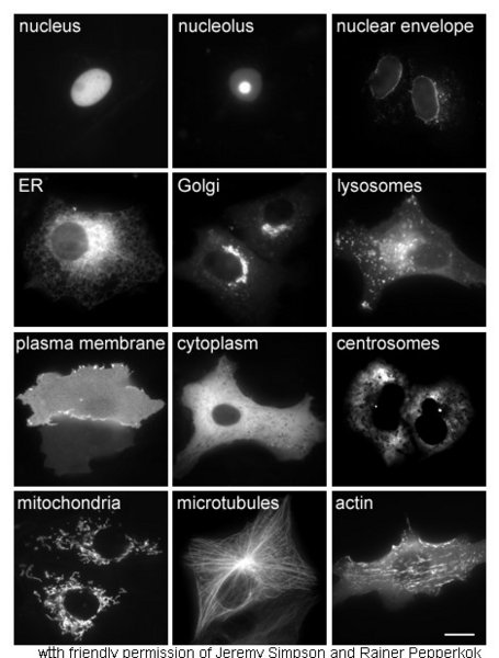 Proteins in different cellular compartments and structures tagged with green fluorescent protein.

References
- ↑ Mader, Sylvia. (2007). Biology 9th ed. McGraw Hill. New York. ISBN 978-0072464634
External links
- Template:McGrawHillAnimation
- 3D structures of proteins associated with lysosome membrane
- Template:NCBI-scienceprimer
ar:جسيمات حالة ca:Lisosoma cs:Lyzozom da:Lysosom de:Lysosom el:Λυσόσωμα eo:Lizosomo gl:Lisosoma ko:리소좀 hr:Lizosom it:Lisosoma he:ליזוזום lb:Lysosom lt:Lizosomos mk:Лизозом nl:Lysosoom no:Lysosom sk:Lyzozóm sl:Lizosom sr:Лизозом sh:Lizozom fi:Lysosomi sv:Lysosom uk:Лізосома