Latissimus dorsi muscle
The latissimus dorsi (plural: latissimi dorsi) is the large, flat, dorso-lateral muscle on the trunk, posterior to the arm, and partly covered by the spinotrapezius on its median dorsal region.
Overview
The latissimus dorsi is a triangular, flat muscle, which covers the lumbar region and the lower half of the thoracic region, and is gradually contracted into a narrow fasciculus at its insertion into the humerus.
It arises by tendinous fibers from the spinous processes of the lower six thoracic vertebrae and from the posterior layer of the lumbodorsal fascia, by which it is attached to the spines of the lumbar and sacral vertebræ, to the supraspinal ligament, and to the posterior part of the crest of the ilium.
It also arises by muscular fibers from the external lip of the crest of the ilium lateral to the margin of the sacrospinalis, and from the three or four lower ribs by fleshy digitations, which are interposed between similar processes of the obliquus abdominis externus.
From this extensive origin the fibers pass in different directions, the upper ones horizontally, the middle obliquely upward, and the lower vertically upward, so as to converge and form a thick fasciculus, which crosses the inferior angle of the scapula, and usually receives a few fibers from it.
The muscle curves around the lower border of the teres major, and is twisted upon itself, so that the superior fibers become at first posterior and then inferior, and the vertical fibers at first anterior and then superior.
It ends in a quadrilateral tendon, about 7 cm. long, which passes in front of the tendon of the teres major, and is inserted into the bottom of the intertubercular groove of the humerus; its insertion extends higher on the humerus than that of the tendon of the pectoralis major.
The lower border of its tendon is united with that of the teres major, the surfaces of the two being separated near their insertions by a bursa; another bursa is sometimes interposed between the muscle and the inferior angle of the scapula.
The tendon of the muscle gives off an expansion to the deep fascia of the arm.
Actions
The latissimus dorsi is responsible for extension, adduction, transverse extension, and internal rotation of the shoulder joint. It also has a synergistic role in extension and lateral flexion of the lumbar spine.
Variations
The number of dorsal vertebræ to which it is attached vary from four to eight; the number of costal attachments varies; muscle fibers may or may not reach the crest of the ilium.
A muscular slip, the axillary arch, varying from 7 to 10 cm. in length, and from 5 to 15 mm. in breadth, occasionally springs from the upper edge of the latissimus dorsi about the middle of the posterior fold of the axilla, and crosses the axilla in front of the axillary vessels and nerves, to join the under surface of the tendon of the pectoralis major, the coracobrachialis, or the fascia over the biceps brachii. This axillary arch crosses the axillary artery, just above the spot usually selected for the application of a ligature, and may mislead the surgeon during the operation. It is present in about 7% of subjects and may be easily recognized by the transverse direction of its fibers.
A fibrous slip usually passes from the lower border of the tendon of the Latissimus dorsi, near its insertion, to the long head of the triceps brachii. This is occasionally muscular, and is the representative of the dorsoepitrochlearis brachii of apes.
Triangles
- The lateral margin of the latissimus dorsi is separated below from the obliquus externus abdominis by a small triangular interval, the lumbar triangle of Petit, the base of which is formed by the iliac crest, and its floor by the obliquus internus abdominis.
- Another triangle is situated behind the scapula. It is bounded above by the trapezius, below by the latissimus dorsi, and laterally by the vertebral border of the scapula; the floor is partly formed by the rhomboideus major. If the scapula is drawn forward by folding the arms across the chest, and the trunk bent forward, parts of the sixth and seventh ribs and the interspace between them become subcutaneous and available for auscultation. The space is therefore known as the triangle of auscultation.
Nerves
The Latissimus dorsi is supplied by the sixth, seventh, and eighth cervical nerves through the thoracodorsal (long subscapular) nerve.
Training
To increase the power of this muscle, the muscle can be trained with the following exercises:
- pull-down (compound)
- bent-over row (compound)
- chin-up and pull-up (compound)
- pull-over (single-joint)
- dumbbell-rows (compound)
- Deadlift (compound)
- T-bar Row (compound)
Most latissimus dorsi exercises concurrently recruit the teres major, posterior fibers of the deltoid, long head of the triceps brachii, among numerous other stabilizing muscles. Compound exercises for the 'lats' typically involve elbow flexion and tend to recruit the biceps brachii, brachialis, and brachioradialis for this function. Depending on the line of pull, the trapezius muscles can be recruited as well; horizontal pulling motions such as rows recruit both latissimus dorsi and trapezius heavily.
Additional images
-
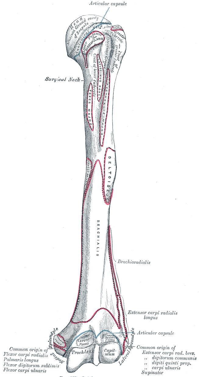 Left humerus. Anterior view.
 -
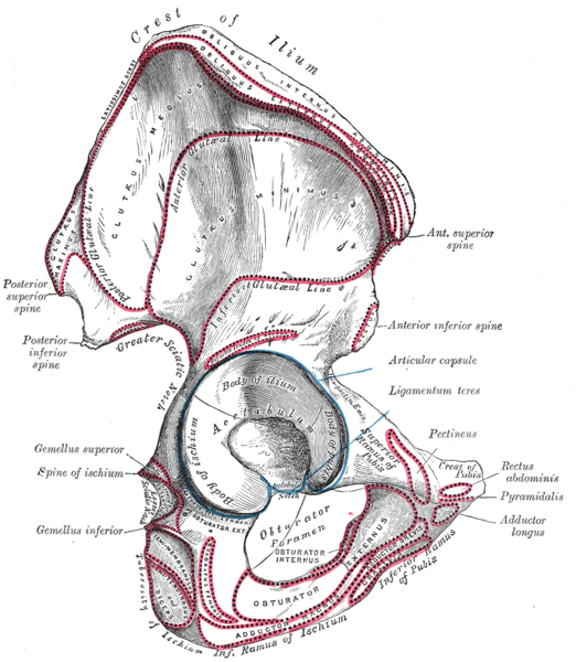 Right hip bone. External surface.
 -
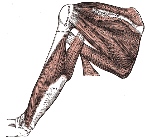 Muscles on the dorsum of the scapula, and the Triceps brachii.
 -
 The axillary artery and its branches.
 -
 The brachial artery.
 -
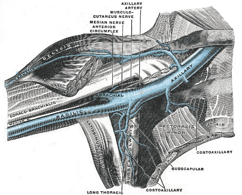 The veins of the right axilla, viewed from in front.
 -
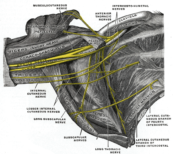 The right brachial plexus (infraclavicular portion) in the axillary fossa; viewed from below and in front.
 -
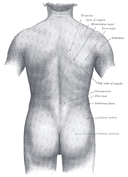 Surface anatomy of the back.
 -
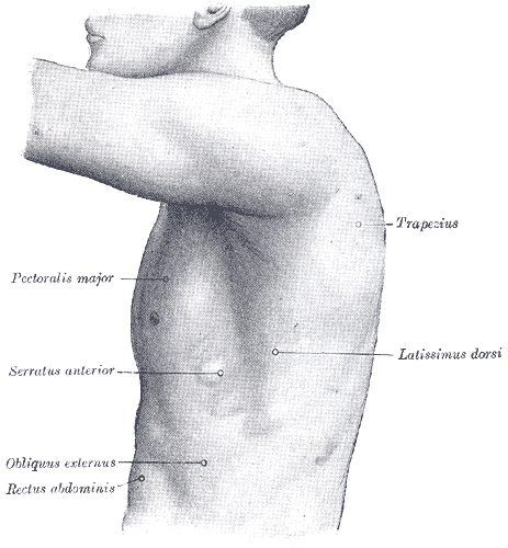 The left side of the thorax.
 -
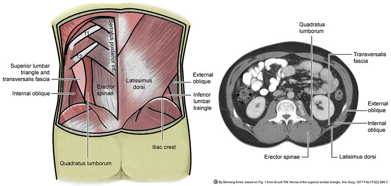 Lumbar triangle


External links
- Template:MuscleLoyola
- Template:GPnotebook
- Template:SUNYAnatomyFigs - "Superficial layer of the extrinsic muscles of the back."
- Template:ViennaCrossSection
- Overview with many illustrations (in context of surgery) at microsurgeon.org
- Illustration at bodybuilding.com
Template:Muscles of upper limb
de:Musculus latissimus dorsi id:Otot latissimus dorsi he:שריר הגב הרחיב nl:Musculus latissimus dorsi sk:Najširší sval chrbta fi:Leveä selkälihas sv:Latissimus dorsi