Fallopian tubes
|
WikiDoc Resources for Fallopian tubes |
|
Articles |
|---|
|
Most recent articles on Fallopian tubes Most cited articles on Fallopian tubes |
|
Media |
|
Powerpoint slides on Fallopian tubes |
|
Evidence Based Medicine |
|
Clinical Trials |
|
Ongoing Trials on Fallopian tubes at Clinical Trials.gov Trial results on Fallopian tubes Clinical Trials on Fallopian tubes at Google
|
|
Guidelines / Policies / Govt |
|
US National Guidelines Clearinghouse on Fallopian tubes NICE Guidance on Fallopian tubes
|
|
Books |
|
News |
|
Commentary |
|
Definitions |
|
Patient Resources / Community |
|
Patient resources on Fallopian tubes Discussion groups on Fallopian tubes Patient Handouts on Fallopian tubes Directions to Hospitals Treating Fallopian tubes Risk calculators and risk factors for Fallopian tubes
|
|
Healthcare Provider Resources |
|
Causes & Risk Factors for Fallopian tubes |
|
Continuing Medical Education (CME) |
|
International |
|
|
|
Business |
|
Experimental / Informatics |
The Fallopian tubes, also known as oviducts, uterine tubes, and salpinges (singular salpinx) are two very fine tubes leading from the ovaries of female mammals into the uterus.
Anatomy
There are two Fallopian tubes, attached to either side of the cornual end of the uterus, and each terminating at or near one ovary forming a structure called the fimbria.
The Fallopian tubes are not directly attached to the ovaries, but open into the peritoneal cavity (essentially the inside of the abdomen); they thus form a direct communication between the peritoneal cavity and the outside via the vagina.
In humans, the Fallopian tubes are about 7–14 cm long.
Regions
There are four regions of the fallopian tube from the ovary to the uterus:[1]
- Infundibulum - contains fimbria
- Ampulla - usual site of fertilization
- Isthmus
- Intramural oviduct - inside wall of uterus
Histology
.JPG)
There are three layers of the fallopian tube:[2]
- Mucosa - the distinctive folds of the mucosa are the most unusual feature. The folds contain ciliated cells and "peg cells".[3][4] The region of the fallopian tube can be approximated by looking at the mucosa, because the folds are most elaborate at the ampulla and almost nonexistent at the intramural portion.
- Muscularis externa
- Serosa
Motility
The Fallopian tubes are mobile, and have been observed on time-lapse videography moving about the pelvis.
Although anatomical illustrations have them proceeding from the uterine horns to the ovary, this is not the case for most of the menstrual cycle, and a tube may cross to the other side or lie on top of the uterus.
Function in fertilization
When an ovum is developing in an ovary, it is encapsulated in a sac known as an ovarian follicle.
On maturity of the ovum, the follicle and the ovary's wall rupture, allowing the ovum to escape and enter the Fallopian tube. There it travels toward the uterus, pushed along by movements of cilia on the inner lining of the tubes. This trip takes hours or days. If the ovum is fertilized while in the Fallopian tube, then it normally implants in the endometrium when it reaches the uterus, which signals the beginning of pregnancy.
Occasionally the embryo implants into the Fallopian tube instead of the uterus, creating an ectopic pregnancy, commonly known as a "tubal pregnancy".
Embryology and homology
The Fallopian tubes are not homologous to the vas deferens or any other structure in males.
Embryos have two pairs of ducts to let gametes out of the body; one pair (the Müllerian ducts) develops in females into the Fallopian tubes, uterus and vagina, while the other pair (the Wolffian ducts) develops in males into the epididymis and vas deferens.
Normally, only one of the pairs of tubes will develop while the other regresses and disappears in the utero.
Pathology
Pelvic inflammatory disease can strike the fallopian tubes. This might cause a fallopian tube obstruction.
Surgery
The surgical removal of a Fallopian tube is called a salpingectomy. To remove both sides is a bilateral salpingectomy. An operation that combines the removal of a Fallopian tube with removal of at least one ovary is a salpingo-oophorectomy. An operation to restore a fallopian tube obstruction is called a tuboplasty.
Etymology and nomenclature
They are named after their discoverer, the 16th century Italian anatomist, Gabriele Falloppio.
Though the name 'Fallopian tube' is eponymous, some texts spell it with a lower case 'f' owing to the theory that the adjective 'fallopian' has been absorbed into modern English as the de facto name for the structure.
The Greek word salpinx (σαλπιγξ) means "trumpet".
Additional images
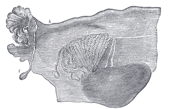
-

Sectional plan of the gravid uterus in the third and fourth month.
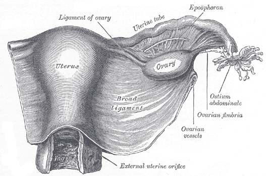
-
Broad ligament of adult, showing epoöphoron.
-
Uterus and right broad ligament,, seen from behind.
-
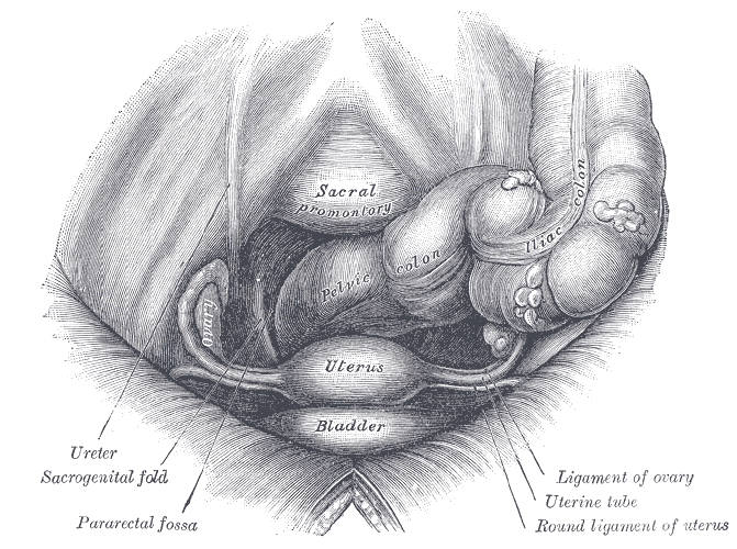
pelvis and its contents, seen from above and in front.
-
Posterior half of uterus and upper part of vagina.
-
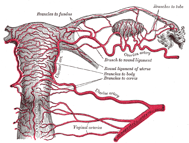
The arteries of the internal organs of generation of the female, seen from behind.
-
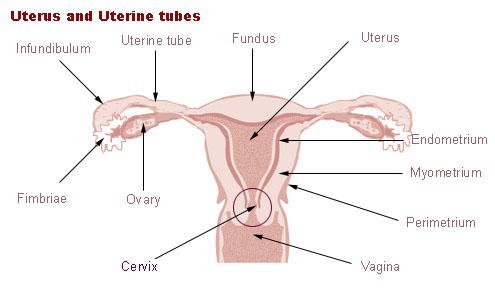
Uterus and uterine tubes.
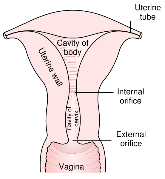
-
Female internal reproductive anatomy.
-
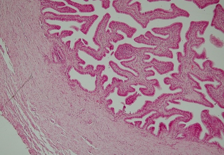
Histology


See also
References
- ↑ Template:SUNYAnatomyLabs - "The Female Pelvis: The Oviduct"
- ↑ Histology at University of Southern California rep/c_27
- ↑ Histology at University of Southern California rep/c_31
- ↑ http://www3.umdnj.edu/histsweb/lab17/lab17oviduct.html
External links
- Template:EMedicineDictionary
- Histology image: 18501loa – Histology Learning System at Boston University
Template:Female reproductive system
ar:قناة فالوب bs:Jajovodi br:Trompilhoù Fallopia bg:Маточна тръба cs:Vejcovod da:Æggeleder de:Eileiter dv:ބިސް ދަތުރުކުރާ ހޮޅި eo:Salpingo id:Tuba fallopi it:Tuba di Falloppio he:חצוצרה (איבר) lt:Kiaušintakis mk:Јајцевод nl:Eileider simple:Fallopian tube sk:Vajíčkovod sl:Jajcevod sr:Јајоводи sh:Jajovodi fi:Munanjohdin sv:Äggledare ta:பாலோப்பியன் குழாய்