Epididymis
Editor-In-Chief: C. Michael Gibson, M.S., M.D. [1]
The epididymis is part of the human male reproductive system and is present in all male mammals. It is a narrow, tightly-coiled tube connecting the efferent ducts from the rear of each testicle to its vas deferens.
Regions
The epididymis can be divided into three main regions
- the head (caput)
- the body (corpus)
- the tail (cauda)
Role in storage of sperm and ejaculation
Spermatozoa formed in the testis enter the caput epididymis, progress to the corpus, and finally reach the cauda region, where they are stored. Sperm entering the caput epididymis are incomplete - they lack the ability to swim forward (motility) and to fertilize an egg. During their transit in the epididymis, sperm undergo maturation processes necessary for them to acquire these functions.[1] Final maturation is completed in the female reproductive tract (capacitation).
During ejaculation, sperm flow from the lower portion of the epididymis (which functions as a storage reservoir). They are packed so tightly that they are unable to swim, but are transported via the peristaltic action of muscle layers within the vas deferens, and are mixed with the diluting fluids of the seminal vesicles and other accessory glands prior to ejaculation (forming semen).
The epididymis is one of only two regions of the body to have stereocilia (the inner ear being the other.)[2]
Pathology
An inflammation of the epididymis is called epididymitis. It is a swollen blood vessel from the testicle that appears or feels like an enlarged epididymis.
Embryology and vestigial structures
A Gartner's duct is a homologous remnant in the female.
Embryologically, the epididymis is derived from tissue that once formed the mesonephros, a primitive kidney found in many aquatic vertebrates. Persistence of the cranial end of the mesonephric duct will leave behind a remnant called the appendix of the epididymis. Additionally, some mesonephric tubules can persist as the paradidymis, a small body caudal to the efferent ductules.
Additional images
-
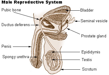 Male reproductive system.
 -
 Testis
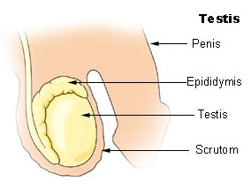
 -
 Schematic drawing of a cross-section through the vaginal process.
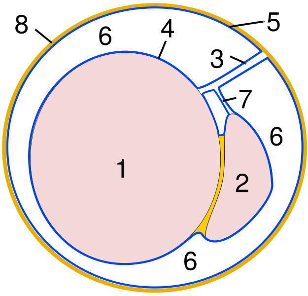
 -
 Microscopic shot.
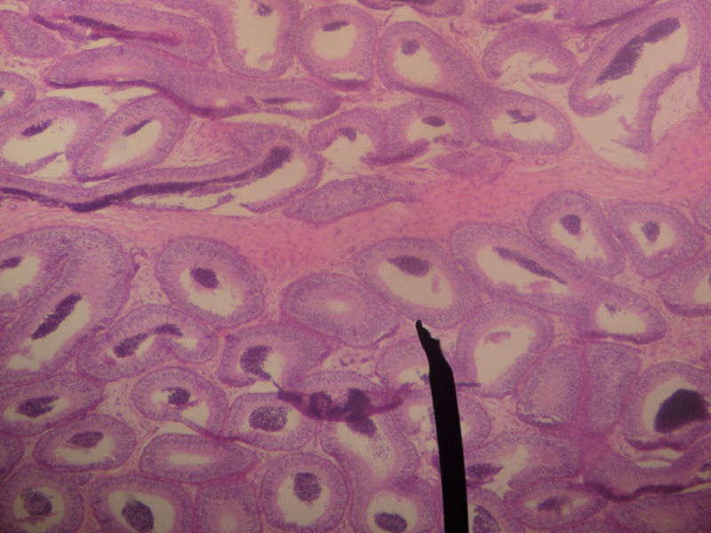


References
- ↑ Jones R (1999). "To store or mature spermatozoa? The primary role of the epididymis". Int J Androl. 22 (2): 57–67. PMID 10194636. abstract
- ↑ http://www.vasectomy-information.com/moreinfo/reabsorb.htm
- Moore, Keith L. & Persaud, T.V.N. (2003). The Developing Human: Clinically Oriented Embryology (7th ed.). Philadelphia: Saunders. ISBN 0-7216-9412-8
External links
- Histology image: 16903loa – Histology Learning System at Boston University
Template:Male reproductive system
ar:بربخ bs:Pasjemenik bg:Надсеменник cs:Nadvarle de:Nebenhoden fa:بربخ it:Epididimo lt:Sėklidės prielipas nl:Bijbal no:Bitestikkel simple:Epididymis sk:Nadsemenník sl:Obmodek fi:Lisäkives sv:Bitestikel