Ovarian follicle
Editor-In-Chief: C. Michael Gibson, M.S., M.D. [1]
Ovarian follicles are the basic unit of female reproductive biology, they are roughly spherical aggregations of cells found in the ovary. They contain a single oocyte (aka ovum or egg). These structures are periodically initiated to grow and develop, culminating in ovulation of usually a single competent oocyte.
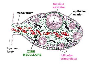
Structure

The cells of the ovarian follicle are the oocyte, granulosa cells and the cells of the internal and external theka layers.
Oocyte
The oocyte in a follicle is in the stage of a primary oocyte. The nucleus of such an oocyte is called a germinal vesicle[1] (see picture).
Granulosa
The oocyte is surrounded by a glycoprotein layer, the zona striata, or zona pellucida. This, in turn, is swaddled in a layer of granulosa cells. In early tertiary follicles, the granulosa cells connecting the oocyte to the rest of the granulosa cells (membrana granulosa are the discus proligerus or cumulus oophorus.
Theka
The granulosa cells, in turn, are enclosed in a thin layer of extracellular matrix – the follicular basement membrane or basal lamina (fibro-vascular coat in picture). Outside the basal lamina, the layers theka interna and theka externa are found.
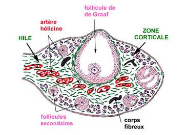
Development

Primordial follocles are undiscernible to the naked eye. However, they develop to primary, secondary and finally mature vesicular follicles. Mature vesicular follicles are sometimes called Graafian follicles (after Regnier de Graaf).
In humans, oocytes are established in the ovary before birth, and may lay dormant awaiting initiation for up to 50 years [2].
After rupturing, the follicle is turned into a corpus luteum.
Development of oocytes in ovarian follicles
In a larger perspective, the whole folliculogenesis from primordial to preovulatory follicle is located in the stage of meiosis I of ootidogenesis in oogenesis.
The embryonic development doesn't differ from the male one, but follows the common path before gametogenesis. Once gametogonia enter the gonadal ridge, however, they attempt to associate with these somatic cells. Development proceeds and the gametogonia turns into oogonia, which become fully surrounded by a layer of cells (pre-granulosa cells).
Oogonia multiply by dividing mitotically; this proliferation ends when the oogonia enter meiosis. The amount of time that oogonia multiply by mitosis is species specific. In the human fetus, cells undergoing mitosis are seen until the second and third trimester of pregnancy [3]; [4]. After beginning the meiotic process, the oogonia (now called primary oocytes) can no longer replicate. Therefore the total number of gametes is established at this time. Once the primary oocytes stop dividing the cells enter a prolonged ‘resting phase’. This ‘resting phase’ or dictyate stage can last anywhere up to fifty years in the human.
For each primary oocyte that undergoes meiosis, only one functional oocyte is produced. The other two or three cells produced are called polar bodies. Polar bodies have no function and eventually deteriorate.
The primary oocyte turns into a secondary oocyte in mature ovarian follicles. Unlike the sperm, the egg is arrested in the secondary stage of meiosis until fertilization.
Upon fertilization by sperm, the secondary oocyte continues the second part of meiosis and becomes a zygote.
Pathology
Any ovarian follicle that is larger than about two centimeters is termed an ovarian cyst.
References
- ↑ Biology-online
- ↑ McGee, E. A., and Hsueh, A. J. (2000). Initial and cyclic recruitment of ovarian follicles. Endocrine Reviews 21, 200-14.
- ↑ Baker, T. G. (1982). Oogenesis and ovulation. In "Book 1: Germ cells and fertilization" (C. R. Austin and R. V. Short, Eds.), pp. 17-45. Cambridge University Press, Cambridge.
- ↑ Byskov, A. G., and Hoyer, P. E. (1988). Embryology of mammalian gonads and ducts. In "The physiology of reproduction" (E. Knobil and J. Neill, Eds.), pp. 265-302. Raven Press, Ltd, New York.
Additional images
-
 Pre-antral follicle
 -
 Graafian follicles


External links
- Template:SUNYAnatomyLabs - "The Female Pelvis: The Ovary"
- Histology image: 14803loa – Histology Learning System at Boston University
- Slide at fda.gov
- Images at okstate.edu
- Life cycle at gfmer.ch
Template:Female reproductive system
de:Ovarialfollikel lt:Grafo folikulas fi:Munarakkula sv:äggblåsa