Brachiocephalic artery
Editor-In-Chief: C. Michael Gibson, M.S., M.D. [1]
The brachiocephalic artery (or brachiocephalic trunk or innominate artery) is an artery of the mediastinum that supplies blood to the right arm and the head and neck.
It is the first branch of the aortic arch, and soon after it is emerges, the brachiocephalic artery divides into the right common carotid artery and the right subclavian artery.
There is no brachiocephalic artery for the left side of the body. The left common carotid, and the left subclavian artery, come directly off the aortic arch. However, there are two brachiocephalic veins.
Origin
It arises, on a level with the upper border of the second right costal cartilage, from the commencement of the arch of the aorta, on a plane anterior to the origin of the left carotid; it ascends obliquely upward, backward, and to the right to the level of the upper border of the right sternoclavicular articulation, where it divides into the right common carotid and right subclavian arteries.
Relations
Anteriorly, it is separated from the manubrium sterni by the Sternohyoideus and Sternothyreoideus, the remains of the thymus, the left innominate and right inferior thyroid veins which cross its root, and sometimes the superior cardiac branches of the right vagus. Posterior to it is the trachea, which it crosses obliquely.
On the right side are the right innominate vein, the superior vena cava, the right phrenic nerve, and the pleura; and on the left side, the remains of the thymus, the origin of the left common carotid artery, the inferior thyroid veins, and the trachea.
Branches
The innominate artery usually gives off no branches, but occasionally a small branch, the thyreoidea ima, arises from it. Other times, it gives off a thymic or bronchial branch.
The thyreoidea ima (a. thyreoidea ima) ascends in front of the trachea to the lower part of the thyroid gland, which it supplies.
It varies greatly in size, and appears to compensate for deficiency or absence of one of the other thyroid vessels. It occasionally arises from the aorta, the right common carotid, the subclavian or the internal mammary.
Additional images
-
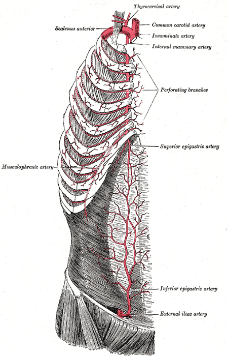 Innominate artery labeled at upper right.
 -
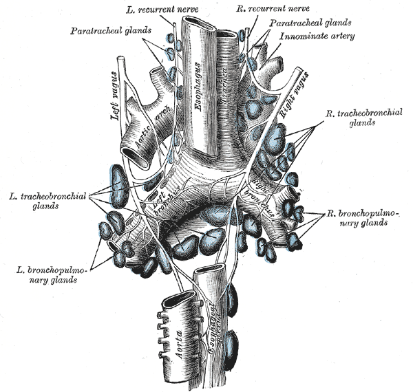 The tracheobronchial lymph glands.
 -
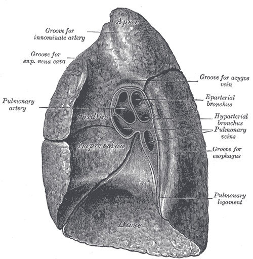 Mediastinal surface of right lung.
 -
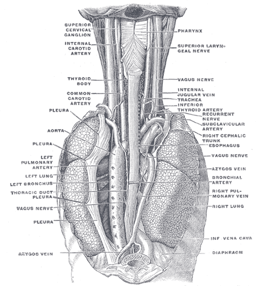 The position and relation of the esophagus in the cervical region and in the posterior mediastinum. Seen from behind.


