Warthin's tumor arises from salivary gland epithelium, which are secretory cells of the salivary gland. On gross pathology, cystic and multicentric appearance are characteristic findings of Warthin's tumor. On microscopic histopathological analysis, papillae, fibrous capsule, and cystic spaces are characteristic findings of Warthin's tumor.
Pathogenesis
Warthin tumor (or papillary cystadenoma lymphomatous) is a benign, sharply demarcated tumor of the salivary gland. It is bilateral in 10-15% of cases.
The first symptom is usually a painless, slow-growing bump in front of the ear, on the bottom of the mouth, or under the chin.
The tumor is slow growing, painless, and usually appears in the tail of the parotid gland near the angle of the mandible.
In 5–14% of cases, Warthin's tumor is bilateral, but the two masses usually are at different times.
Warthin's tumor is highly unlikely to become malignant.[1][2]
Genetics
Expression of CRTC1 - MAML2 chimeric genes through t(11;19)(q21;p13) translocation is involved in the pathogenesis of Warthin's tumor.
Gross Pathology
Warthin's tumor is often multicentric (20%) and is usually small (1-4 cm). It has typically heterogeneous appearance on all modalities, often with cystic components (30%).[3][4]
The size of Warthin's tumor is usually 2.5 centimeter.
Warthin's tumor tend to favor the parotid tail region.
Microscopic Pathology
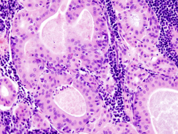
The appearance of this tumor under the microscope is unique. There are cystic spaces surrounded by two uniform rows of cells with centrally placed pyknotic nuclei.
The cystic spaces have epithelium referred to as papillary infoldings that protude into them. Additionally, the epithelium has lymphoid stroma with germinal center formation.
Papillae (nipple-shaped structures) with a two rows of pink (eosinophilic) epithelial cells (with cuboidal basal cells and columnar luminal cells) - key feature
Fibrous capsule - pink & homogenous on H&E stain
Cystic space filled with debris in situ (not necrosis)
↑ 6.06.1Abid, Syed A.; Stack, Brendan C.; Bodenner, Donald L. (2014). "Metastatic Follicular Thyroid Carcinoma Secreting Thyroid Hormone and Radioiodine Avid without Stimulation: A Case Report and Literature Review". Case Reports in Endocrinology. 2014: 1–6. doi:10.1155/2014/584513. ISSN2090-6501.
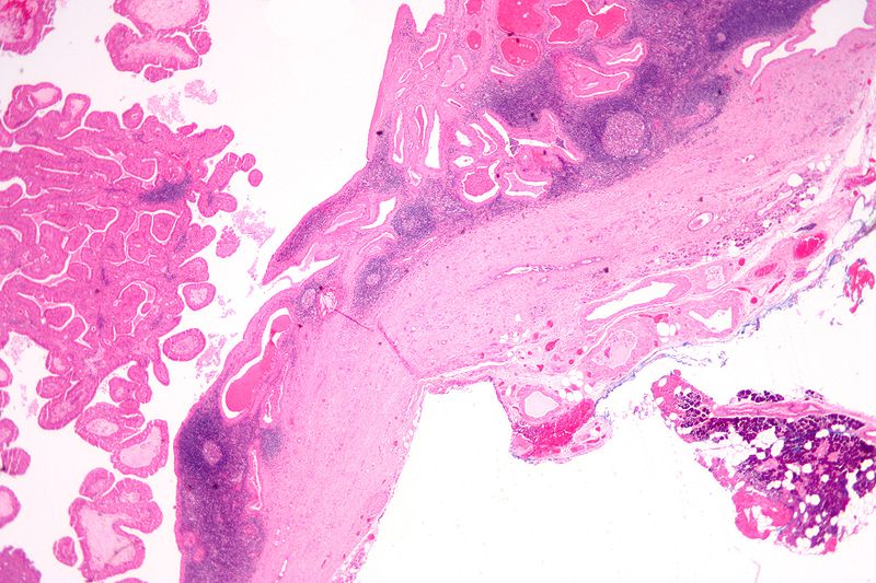
 Histopathology of Warthin tumor in the parotid gland. H&E stain Source:<ref name
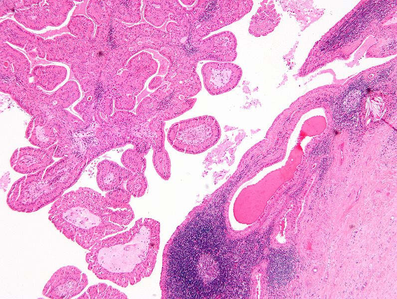
![Histopathology of Warthin tumor in the parotid gland. Another view of a file "Warthin tumor (1).jpg". H&E stain..[6]](/images/c/cb/Warthin_tumor_%282%29.jpg)
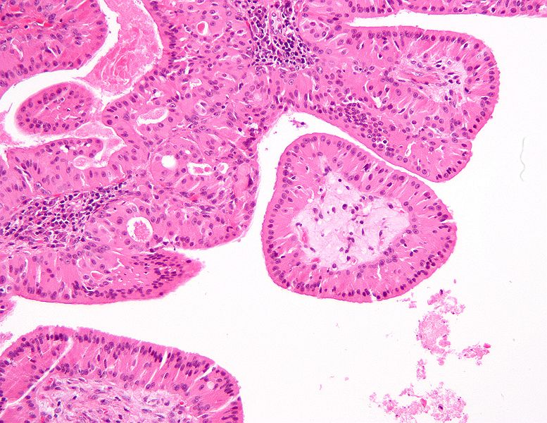
![Histopathology of Warthin tumor in the parotid gland. Higher magnification of a file "Warthin tumor (1).jpg". H&E stain.[6]](/images/d/d2/Warthin_tumor_%283%29.jpg)
.jpg)
![Histopathology of Warthin tumor in the parotid gland. Another view of a file "Warthin tumor (1).jpg". H&E stain..[6]](/index.php/File:Warthin_tumor_(2).jpg)
![Histopathology of Warthin tumor in the parotid gland. Higher magnification of a file "Warthin tumor (1).jpg". H&E stain.[6]](/index.php/File:Warthin_tumor_(3).jpg)


