Gallery of new files
Jump to navigation
Jump to search
This special page shows the last uploaded files.
-
-
-
-
-
-
-
-
-
-
-

-
-
-

-
-
-
-
-
-
-
-
-

-
-
-

-
-
-
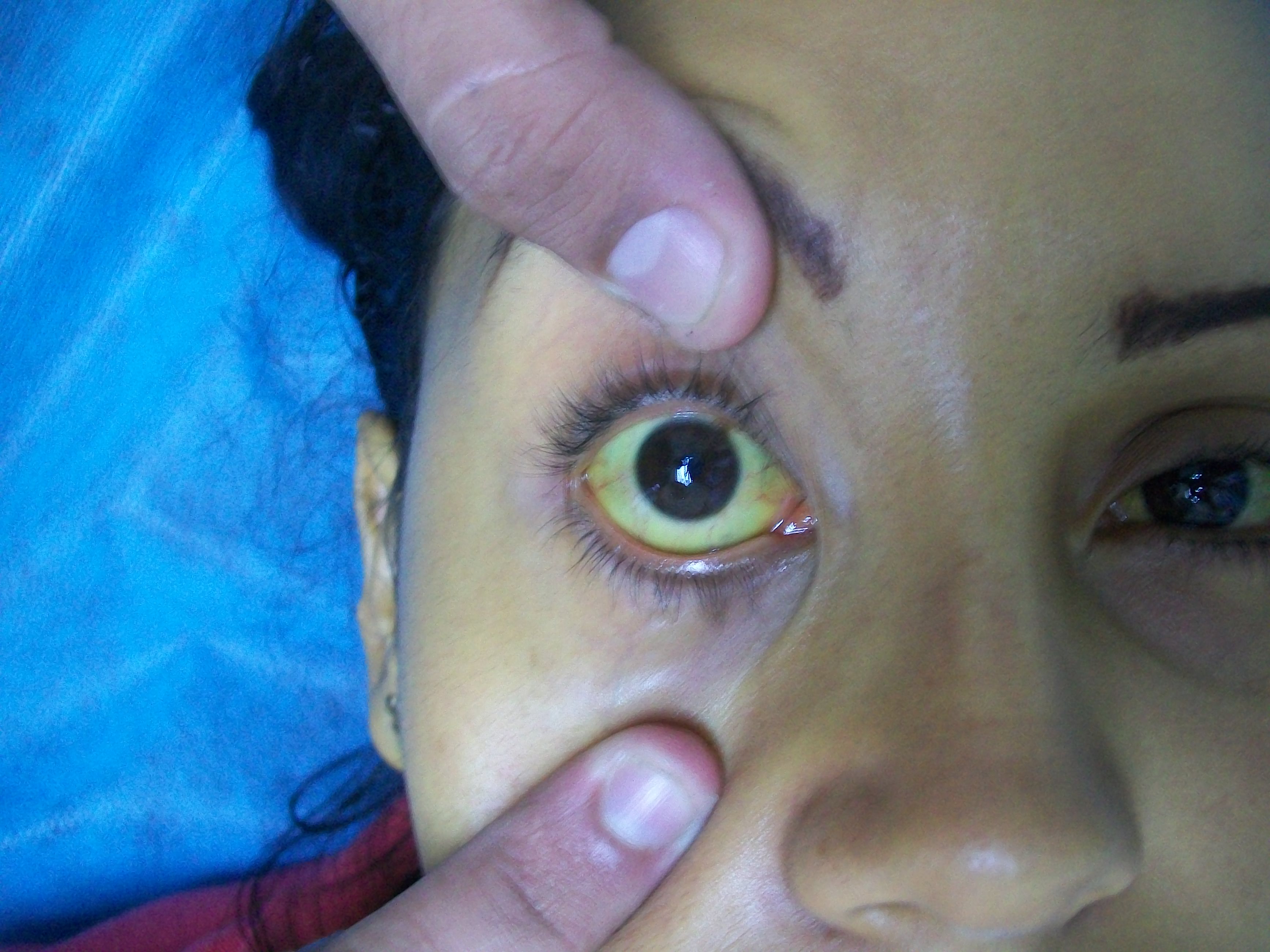
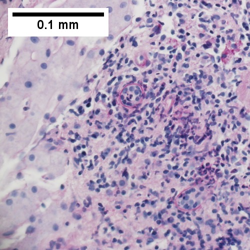
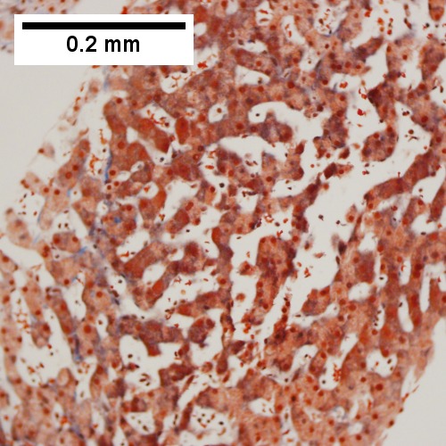
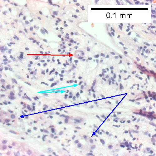
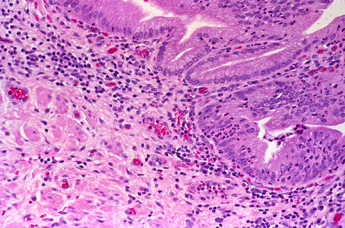
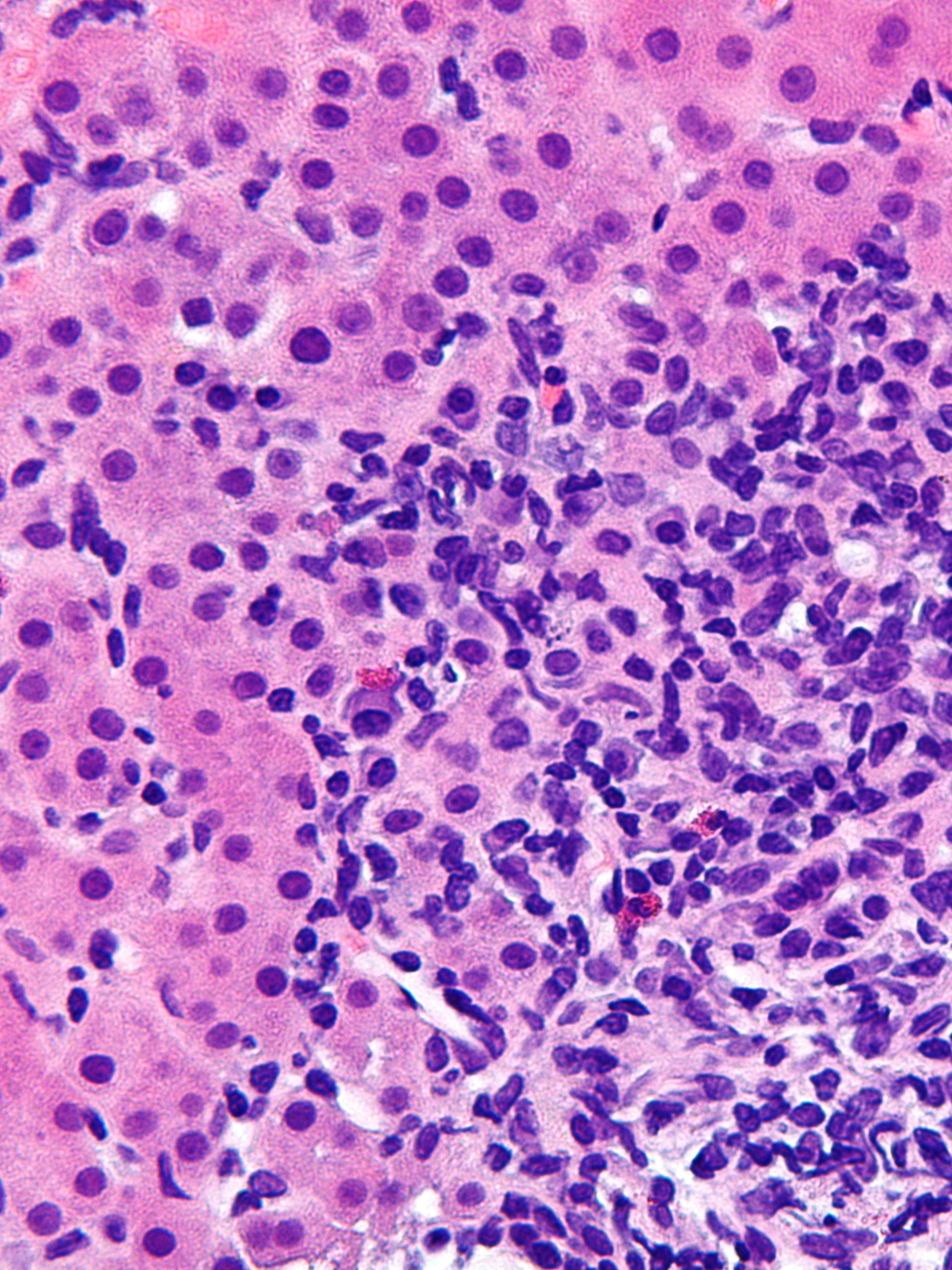 Autoimmune hepatitis - cropped - very high mag.jpg Manpreet Kaur
20:15, 26 December 2017
2,136 × 2,848; 2.74 MB
-
-
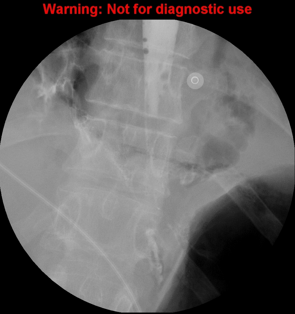
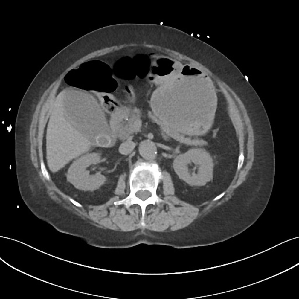
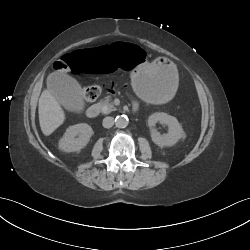
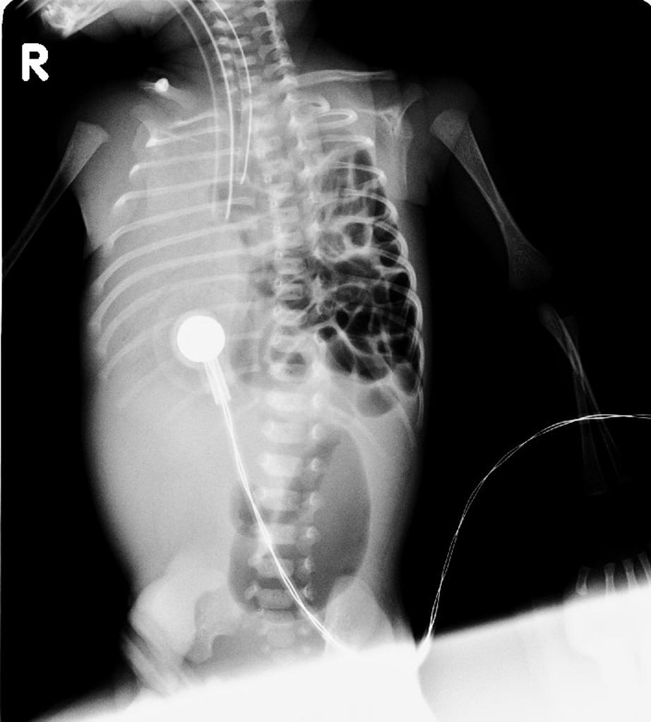
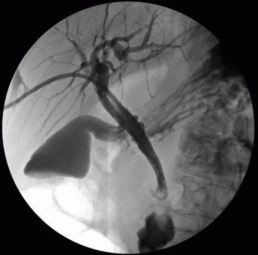
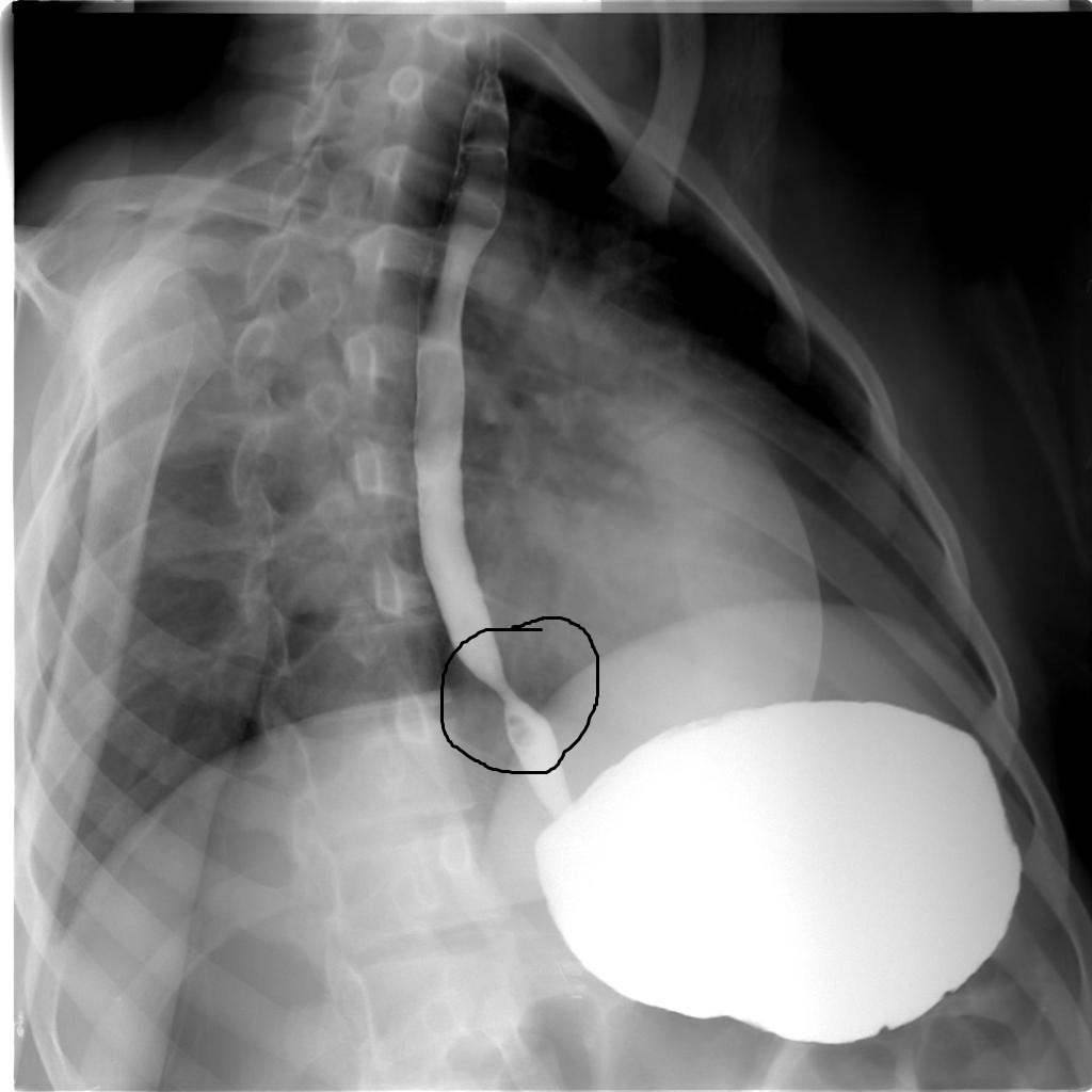
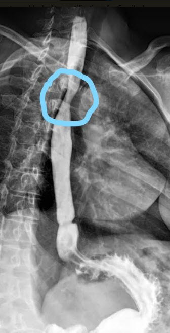
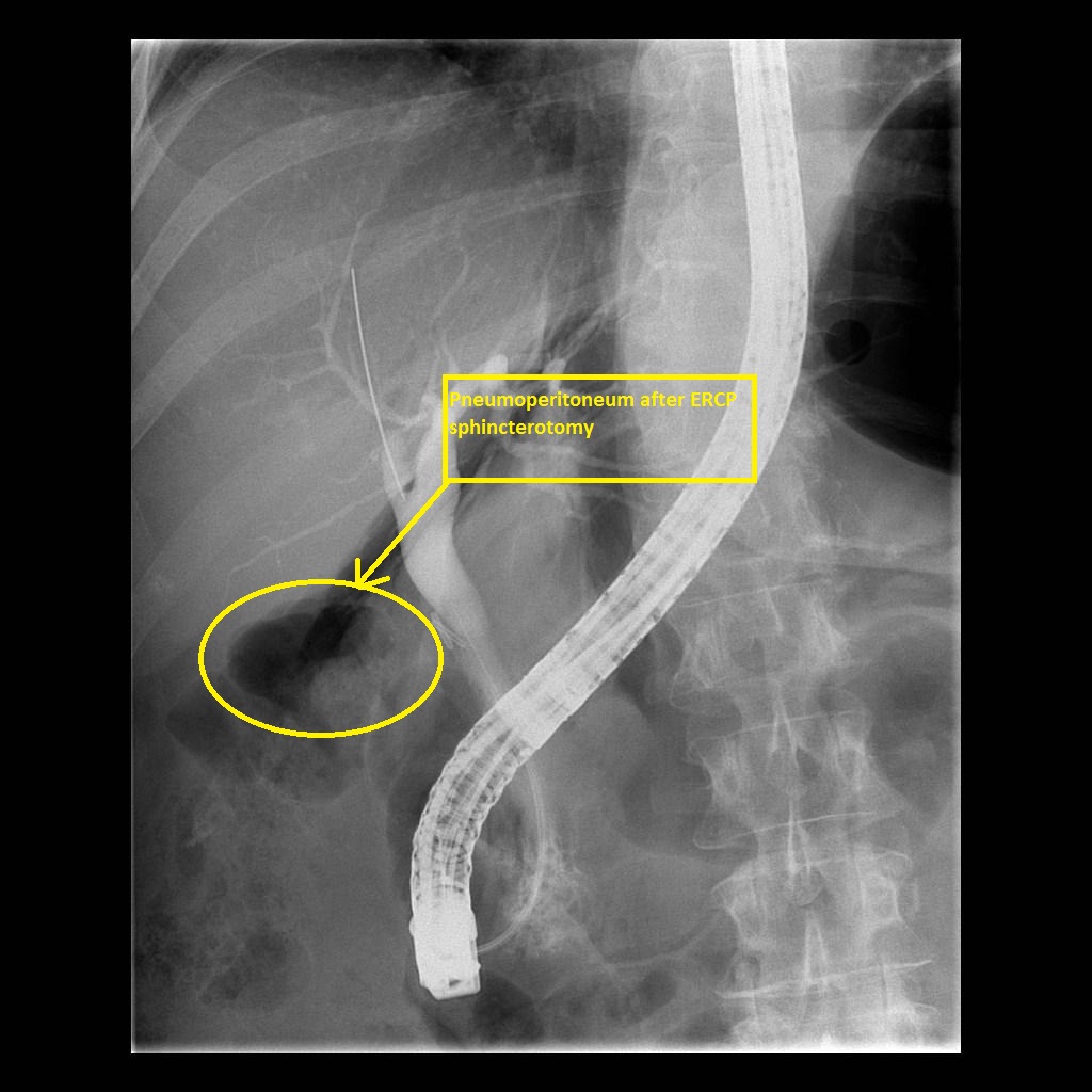 Pneumoretroperitoneum-after-ercp-sphincterotomy.jpg Iqra Qamar
17:24, 22 December 2017
1,024 × 1,024; 166 KB
-

-
-
-

-

-
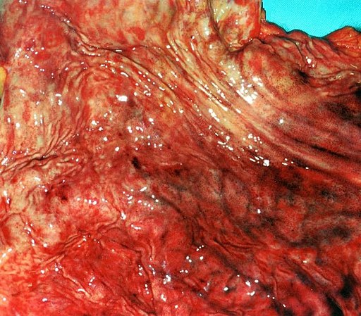
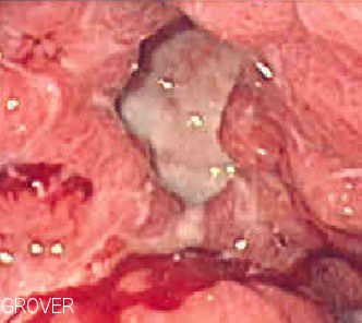
 Multiple polyps and at large mass at the hepatic flexure.jpg Hamid Qazi
17:03, 21 December 2017
960 × 720; 51 KB
-
-
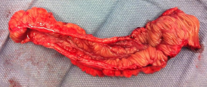
 Image of resected colon segment with cancer & 4 nearby polyps plus schematic of field defects with sub-clones.jpg Ssharfaei
17:12, 20 December 2017
1,672 × 2,560; 625 KB
-
-

-
-
-
-
-
-
-
-
-
-
 Acute cholecystitis - a -- intermed mag.jpg Furqan M Muhammad
18:59, 19 December 2017
320 × 480; 62 KB
-

-
-

-

-
-
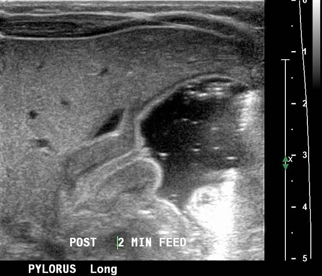
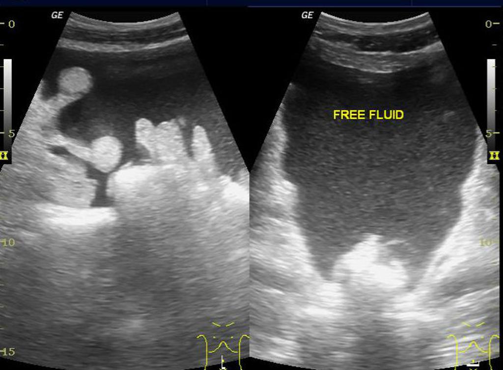
 Ascending-colo-colic-intussusception-due-to-lipoma.jpg Sargun Walia
16:43, 19 December 2017
957 × 1,024; 87 KB
-

-
-

-

-
-
-

-
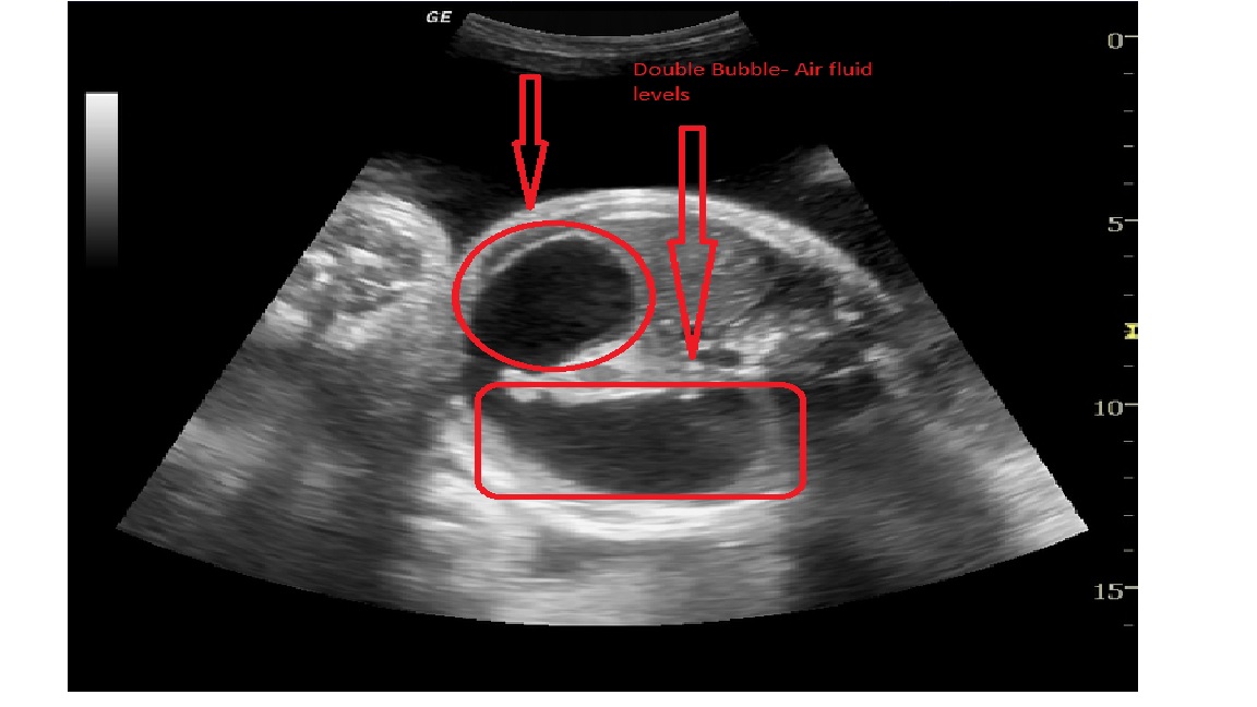
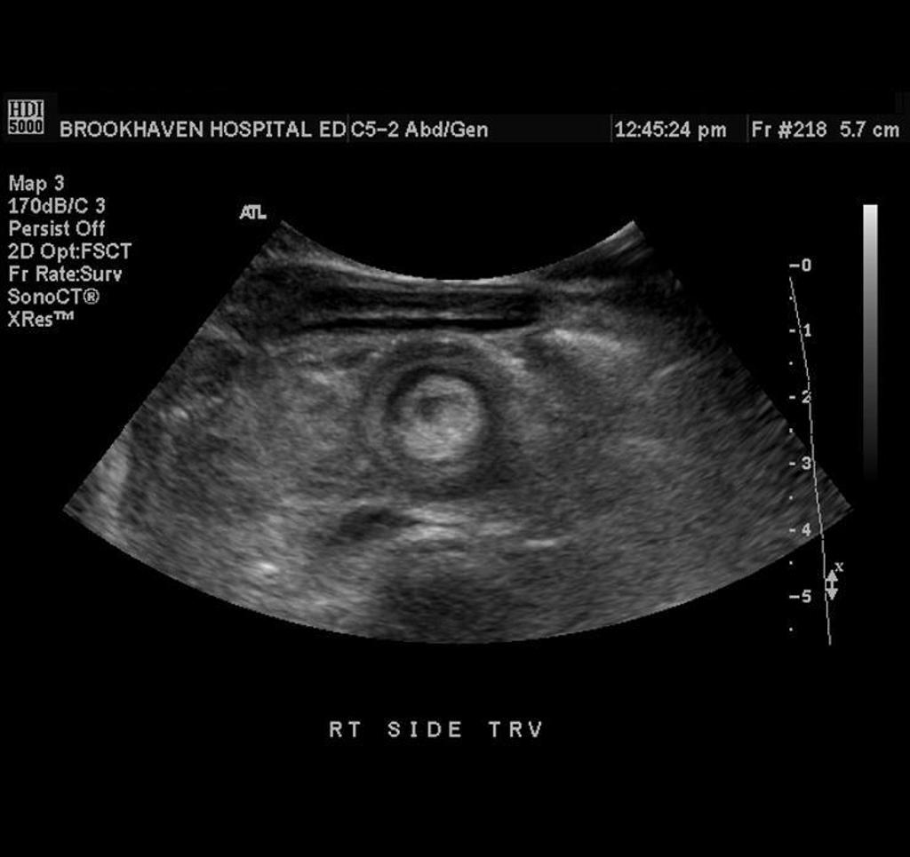
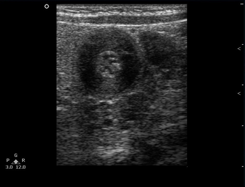
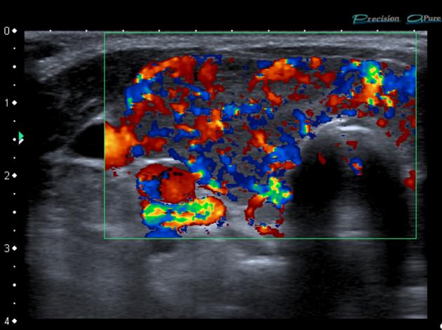
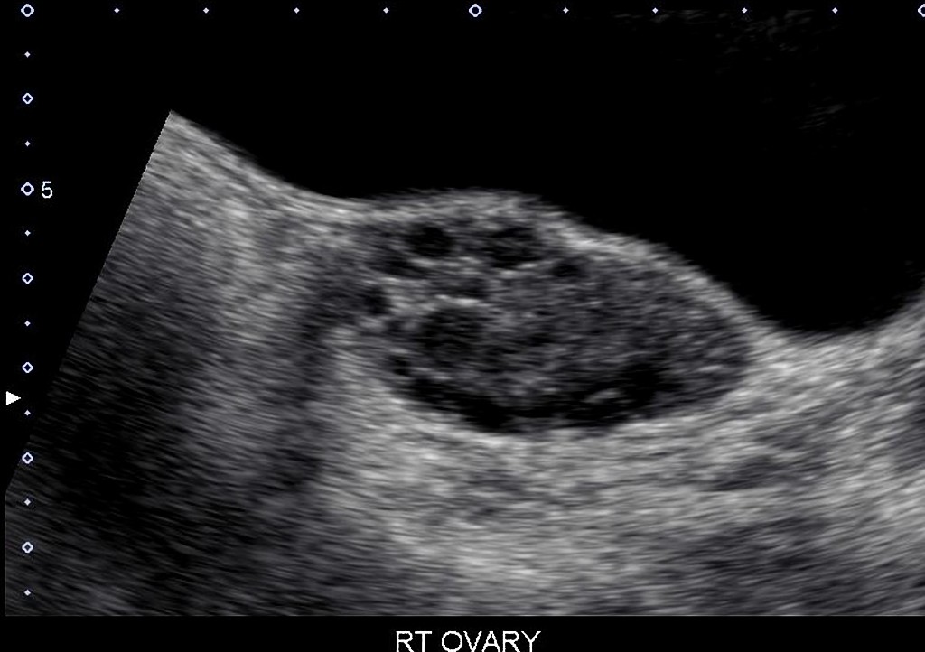
 Intussusception- Transverse view on Ultrasound 2.jpg Sargun Walia
19:36, 18 December 2017
1,024 × 965; 75 KB
-
-

-
-
-

-

-
-
-
 CT image of a GIST tumor in the gastric cardia.jpg Akshun Kalia
23:20, 17 December 2017
682 × 512; 76 KB
-

-

-
-
-

-
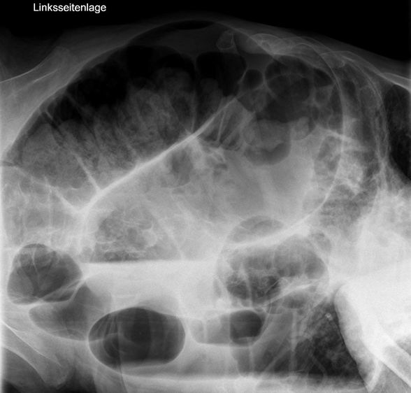
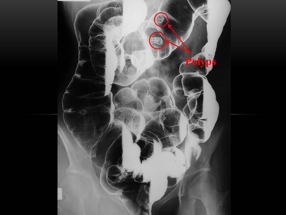
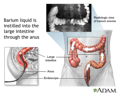
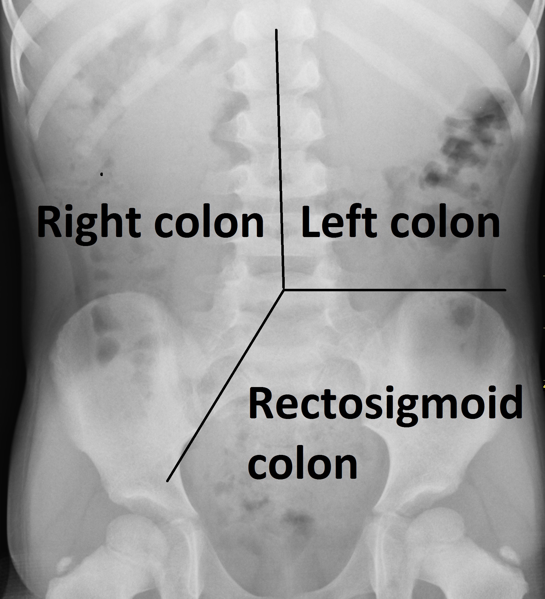
 Human intestinal tract, as imaged via double-contrast barium enema.jpg Eiman
17:30, 15 December 2017
387 × 450; 78 KB
-
-
-

-

-

-

-

-
-

-

-

-

-
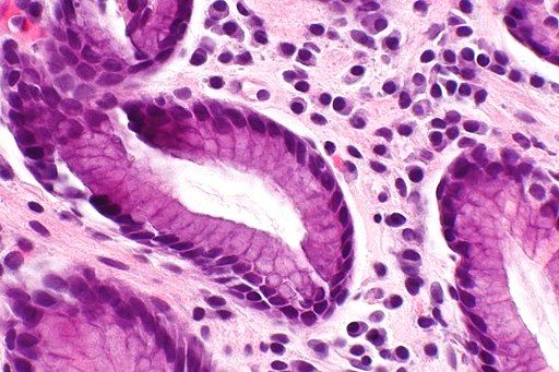 Chronic gastritis -- very high mag.jpg Aravind Reddy Kothagadi
16:38, 14 December 2017
512 × 341; 63 KB
-
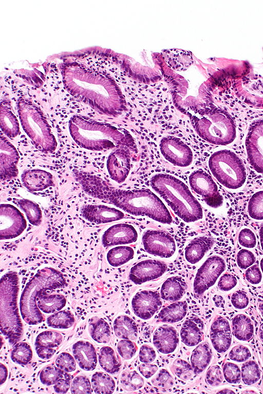 Chronic gastritis -- intermed mag.jpg Aravind Reddy Kothagadi
16:38, 14 December 2017
512 × 768; 155 KB
-

-

-
-
-
-
-

-

-

-
-

-
-

-
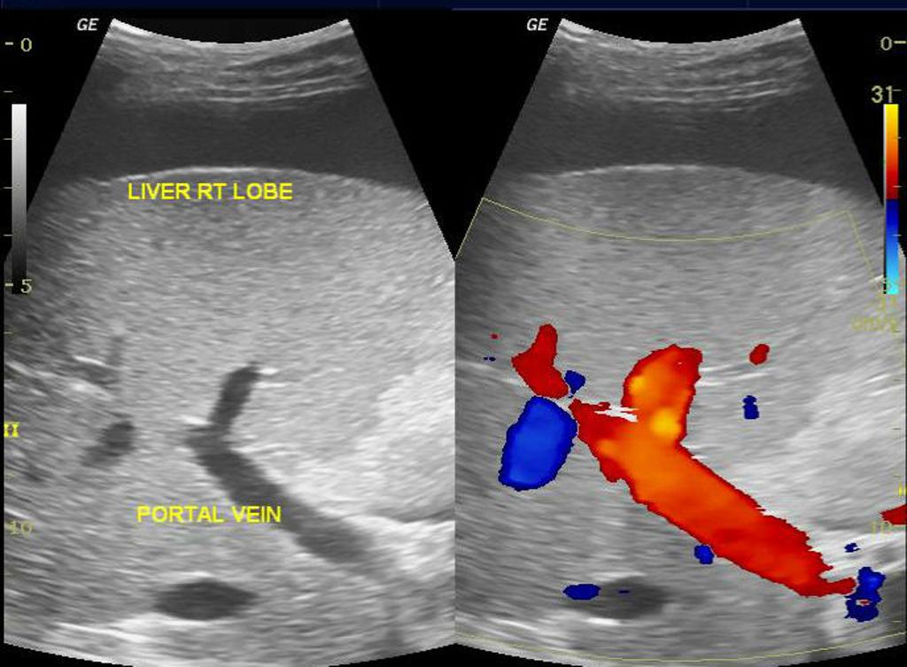
 Cavernous-transformation-of-the-right-portal-vein-branch.jpg Farima Kahe
20:10, 12 December 2017
1,024 × 787; 47 KB
-

-

-

-
-
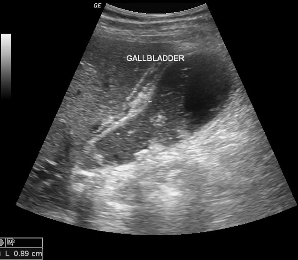
 Acute-cholecystitis-with-gallbladder-neck-calculus (1).jpg Hadeel Maksoud
18:26, 11 December 2017
1,324 × 740; 104 KB
-
 Acute-cholecystitis-with-gallbladder-neck-calculus.jpg Hadeel Maksoud
18:21, 11 December 2017
1,024 × 756; 110 KB
-
-

-

-

-

-

-
-
-
-
-
-
-

-

-
-
-
-
-
-

-
-
-
-

-

-

-

-

-

-

-

-

-

-

-

-

-

-

-

-
-
-
-
-
-
-
-
-
-
-
-
-
-

-
-
-
-

-
-
-

-

-

-

-

-

-

-

-
-
-
-

-

-
-
-
-

-

-

-
-
-

-

-

-

-

-

-

-
-

-

-

-

-

-

-
-

-

-
-
-
-

-

-
-

-
-

-

-

-

-
-

-
-

-

-

-

-
-
-

-
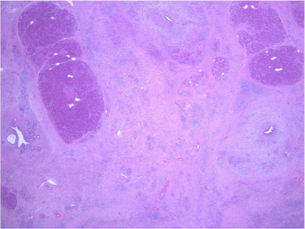
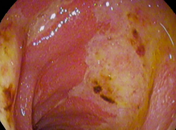
 Adenocarcinoma low differentiated (stomach) H&E magn 400x.jpg Medhat
19:55, 20 November 2017
1,280 × 1,024; 155 KB
-

-

-
-

-

-

-

-

-

-
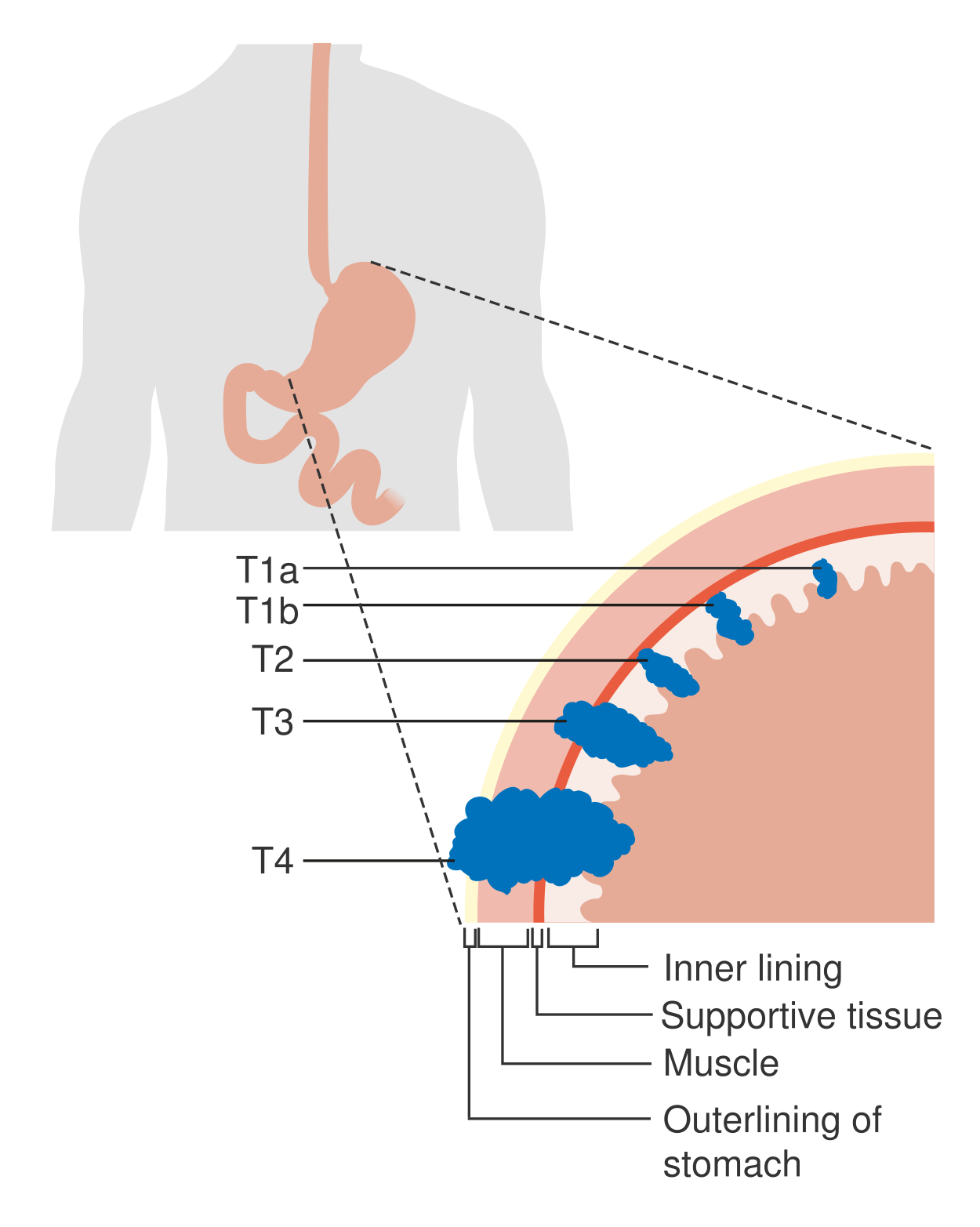 Diagram showing the T stages of stomach cancer CRUK 374.png Medhat
22:20, 16 November 2017
1,250 × 1,565; 124 KB
-

-

-

-

-

-
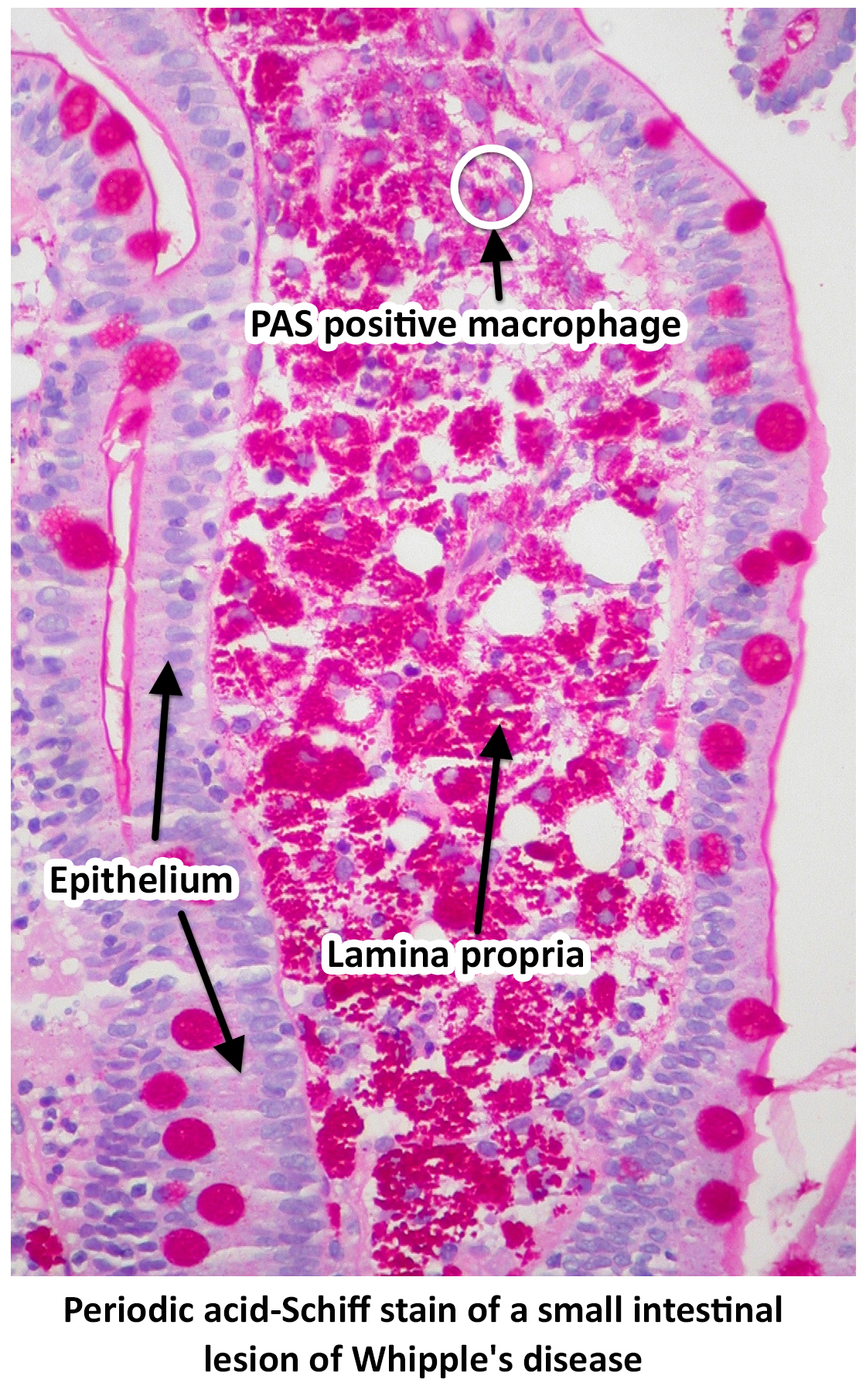
 Light microscopy of intestine-Whipples Disease.jpg .jpg Ssharfaei
15:04, 16 November 2017
762 × 512; 430 KB
-

-

-

-
-

-
-

-

-

-

-

-
-
-
-
-
-
-
-

-

-

-
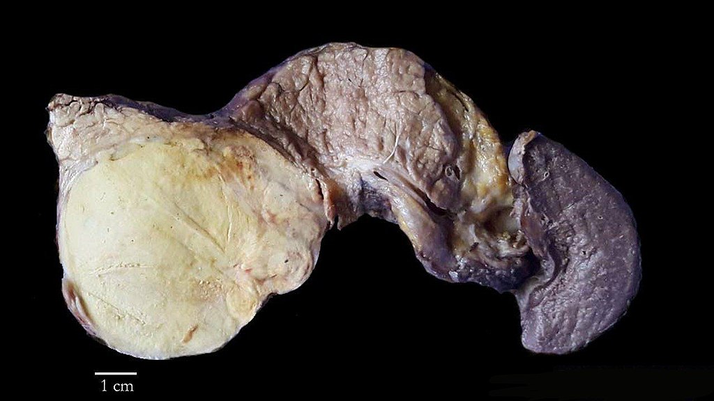
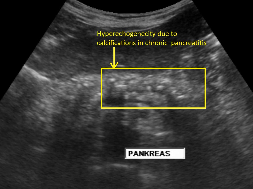 Pancreatic-calcifications-in-chronic-pancreatitis.jpg Iqra Qamar
14:31, 14 November 2017
1,024 × 767; 118 KB
-

-

-
 Light microscopy of intestine-Whipples Disease.jpg Ssharfaei
21:26, 13 November 2017
762 × 512; 422 KB
-
-

-

-

-

-
-

-
 Eingeblutete Pankreaspseudozyste - CTpv cor1 001.JPG Iqra Qamar
16:44, 8 November 2017
1,024 × 1,270; 500 KB
-
 Eingeblutete Pankreaspseudozyste - CTpv axial 001.JPG Iqra Qamar
16:34, 8 November 2017
1,024 × 784; 303 KB
-
 Chronische Pankreatitis mit Verkalkungen - CT axial.jpg Iqra Qamar
16:28, 8 November 2017
1,158 × 956; 223 KB
-

-
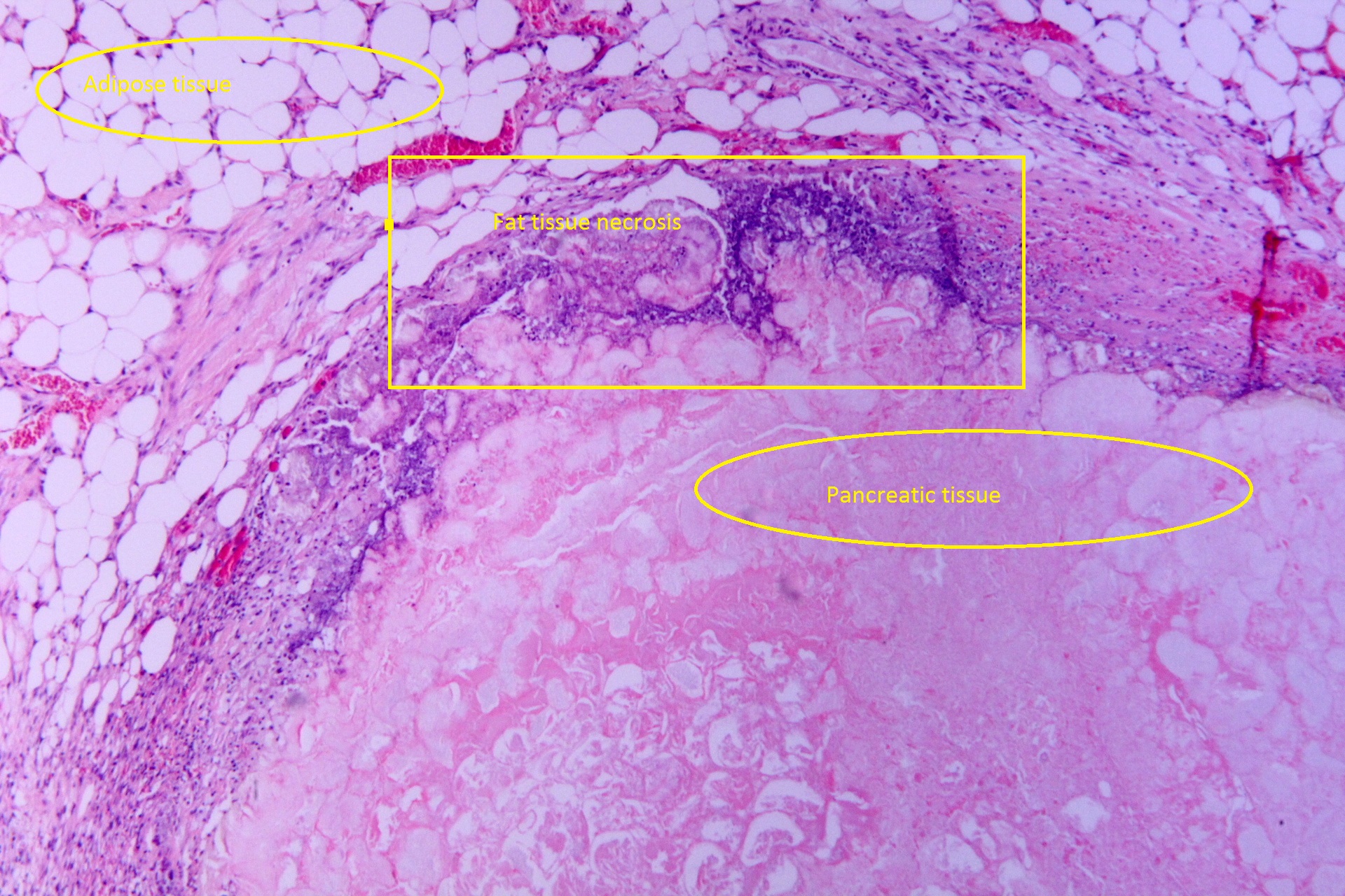
 Tryptic fat tissue necrosis in severe pancreatitis, HE 2.jpg Iqra Qamar
16:05, 8 November 2017
1,920 × 1,280; 1.39 MB
-

-
-

-
 Tryptic fat tissue necrosis in severe pancreatitis, HE 1.jpg Iqra Qamar
15:01, 8 November 2017
1,920 × 1,280; 923 KB
-
 Hemorrhagic pancreatitis - Grey Turner's sign.jpg Iqra Qamar
14:46, 8 November 2017
602 × 316; 27 KB
-

-
-
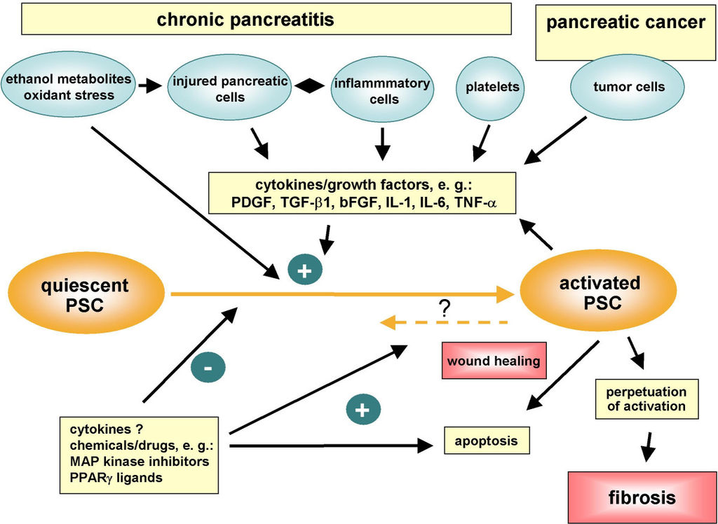 1024px-Pancreatic stellate cell activation in chronic pancreatitis and pancreatic cancer.jpg Iqra Qamar
14:03, 8 November 2017
1,024 × 748; 104 KB
-

-

-

-

-

-

-
-
-
-
-

-
-
-
-
-
-

-
-
-
-
-

-

-

-

-

-
-
 22908713 10214747514538937 676744570 o.jpg Mazia Fatima
16:45, 2 November 2017
1,334 × 1,334; 110 KB
-

-

-

-
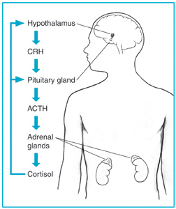
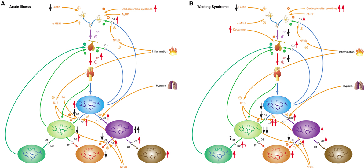 Acute NTIS and Wasting Syndrome.jpeg Johannes W. Dietrich
18:51, 1 November 2017
1,192 × 550; 357 KB
-
-
-
-
-
-
-
-
-

-

-
-

-
-
-

-

-
-

-
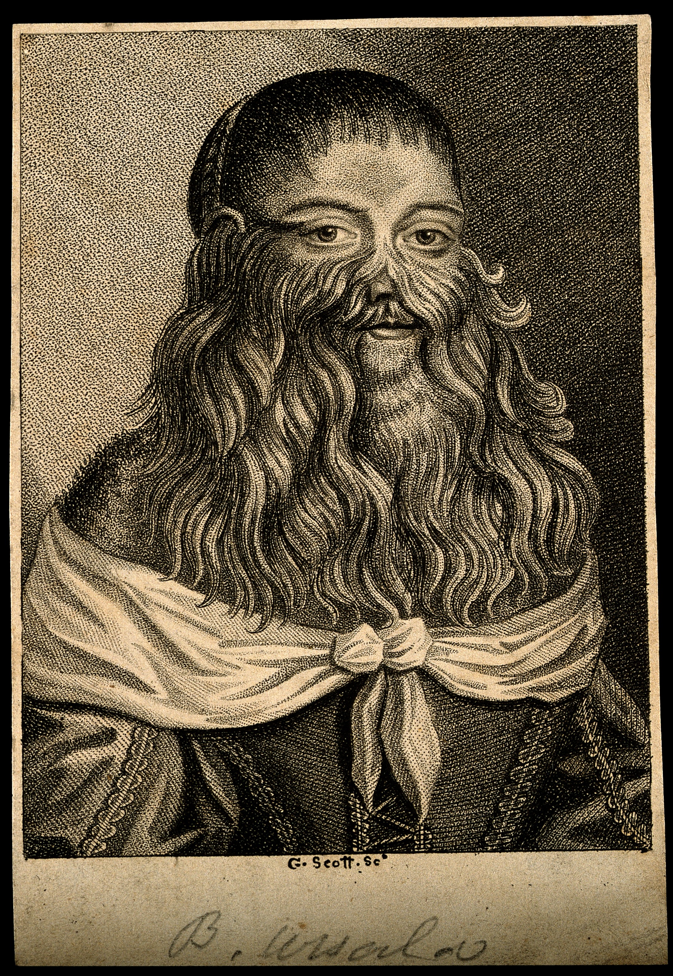
 F. Glisson, plate II,-Anatomia hepatis- Wellcome L0013987.jpg Eiman
17:01, 30 October 2017
1,454 × 1,318; 512 KB
-
 Rene-Theophile-Hyacinthe Laennec (1781-1826) Drawings diseased lungs.jpg Eiman
16:55, 30 October 2017
820 × 800; 197 KB
-

-
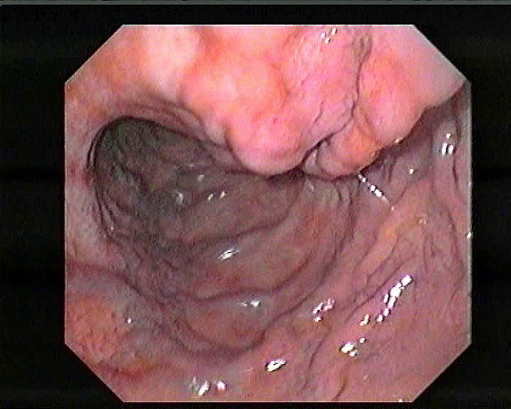
 Gastroesophageal reflux disease -- high mag.jpg Aelsaiey
15:14, 30 October 2017
4,272 × 2,848; 4.45 MB
-

-

-

-
-
-
-

-

-
-
-
-
 F21. Venous enlargement in hepatic cirrhosis. Alfred Kast Wellcome L0074357.jpg Eiman
19:24, 26 October 2017
2,718 × 3,681; 3.68 MB
-
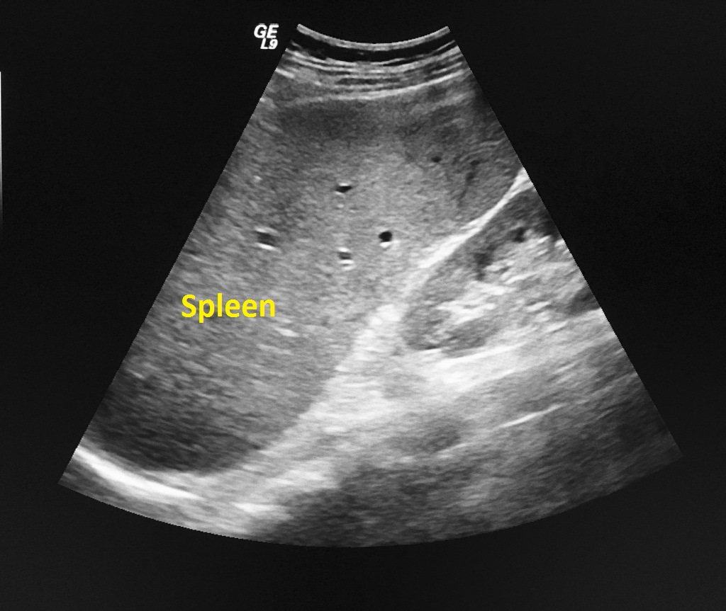
 Esplenomegalia i hiperplasia linfoide folicular reactiva. IMG 2865.jpg Eiman
19:15, 26 October 2017
670 × 453; 90 KB
-

-

-

-

-
-
-

-
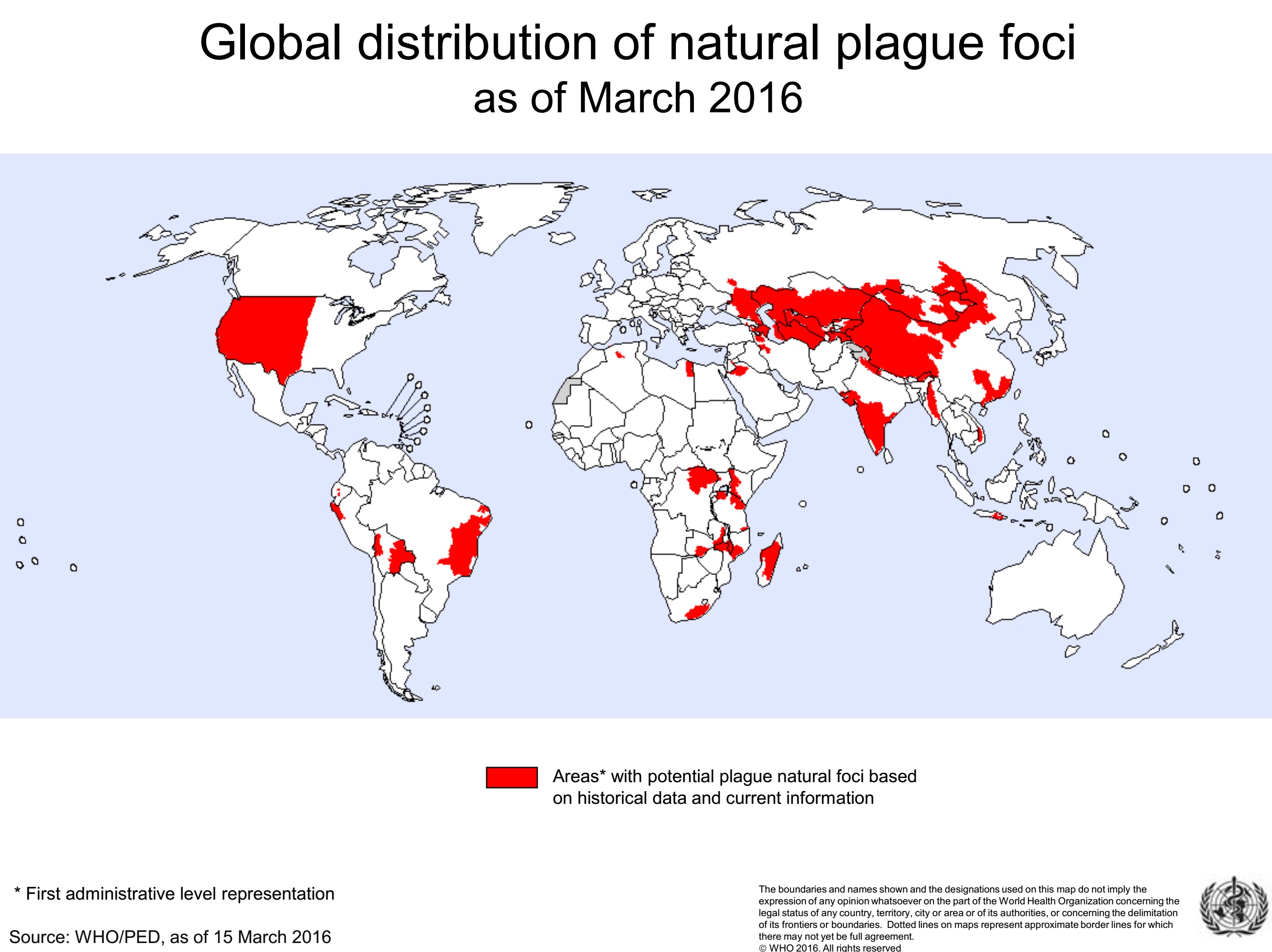 Plague 2016 global distribution.jpg Aravind Reddy Kothagadi
12:13, 24 October 2017
2,695 × 2,018; 601 KB
-
-

-

-
-

-
-

-
-

-
-

-

-
-

-

-

-

-

-

-
-

-

-
-
-
-
-
-
-
-

-
-

-

-

-

-

-

-
-

-
-

-
-
-
-
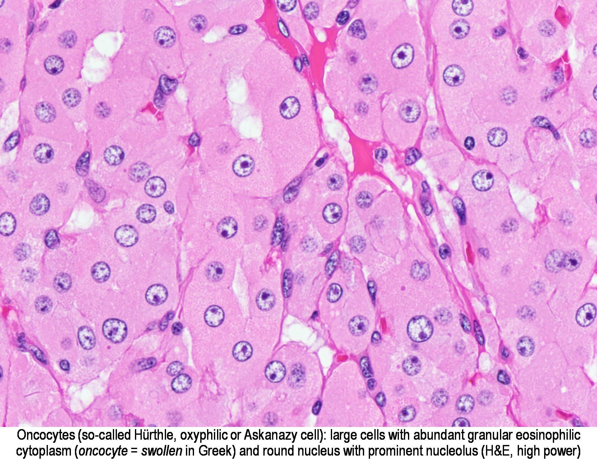
 Hurthle cell adenoma-histology 40x H&E.jpg Furqan M Muhammad
06:46, 7 October 2017
1,024 × 771; 382 KB
-

-
-

-

-

-
-
-
-

-
 Trends in birth prevalence of congenital Cerebral Palsy.jpg Aditya Ganti
16:32, 5 October 2017
500 × 218; 63 KB
-
-

-

-
-
-
-
-

-
-
-

-

-
-
-
-
-

-
-

-

-
-

-

-
-

-
-
-

-
-
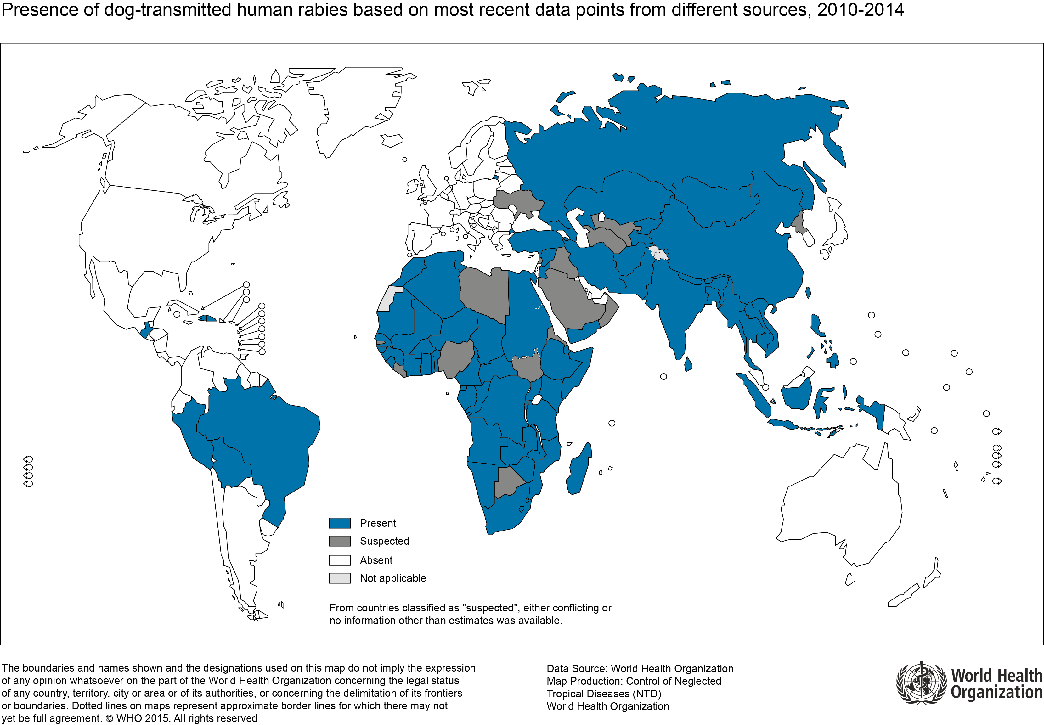 Presence dog transmitted human Rabies 2014.png Skazmi
22:21, 27 September 2017
3,317 × 2,304; 613 KB
-

-
-

-

-

-

-

-

-
-

-
-
-
 Hashimoto-thyroiditis-with-consequent-hypothyroidism.jpg Furqan M Muhammad
16:15, 25 September 2017
1,024 × 710; 121 KB
-

-

-

-
-

-
-
-
-
-

-
-
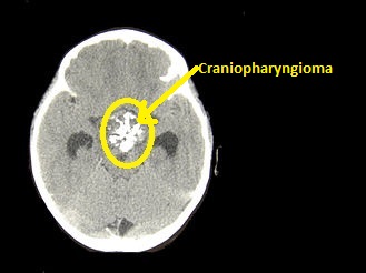
 800px-CNS Craniopharyngeoma AdamantomatousType MP CTR.jpg Eiman
21:48, 21 September 2017
800 × 600; 284 KB
-
-
-

-

-
-
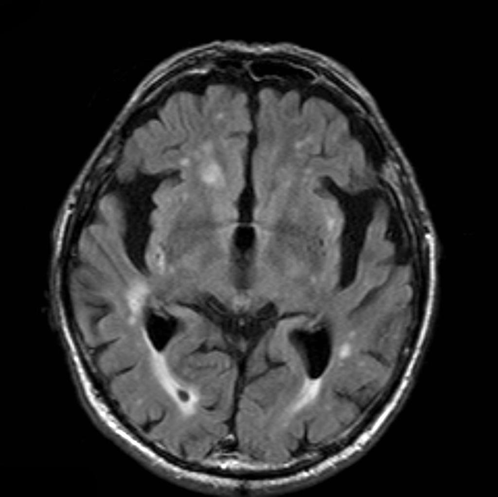
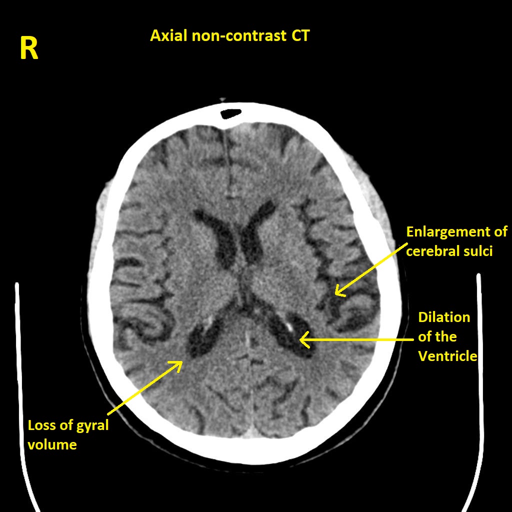 Alzheimers disease axial non contrast CT.jpg Aravind Reddy Kothagadi
18:09, 21 September 2017
1,024 × 1,024; 148 KB
-

-

-
-

-

-
-
-
 NF tangles in the Hippocampus Alzheimer tau protein.JPG Aravind Reddy Kothagadi
01:44, 21 September 2017
1,200 × 800; 214 KB
-

-
 Neurofibrillary tangles in the Hippocampus HE 3.JPG Aravind Reddy Kothagadi
01:38, 21 September 2017
1,920 × 1,280; 1.52 MB
-
 Alzheimer's disease brain comparison.jpg Aravind Reddy Kothagadi
00:24, 21 September 2017
511 × 231; 35 KB
-
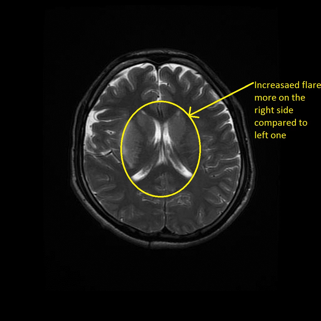
 MRI - alzheimer-disease-FLAIR.jpg Aravind Reddy Kothagadi
20:06, 20 September 2017
1,024 × 1,021; 75 KB
-
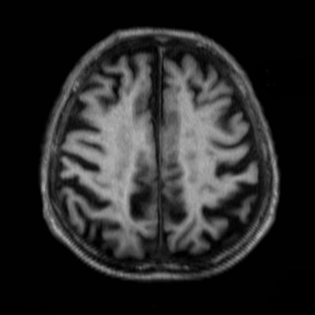 MRI - alzheimer-disease-T1.jpg Aravind Reddy Kothagadi
20:06, 20 September 2017
1,024 × 1,024; 58 KB
-


.jpg)
.jpg)
.jpg)


.jpg)


.jpg)


.jpg)


.gif)


.jpg)

.jpg)


.jpg)


.jpg)


.gif)
.png)


-1.jpg)
_H%26E_magn_400x.jpg)

.jpg)


.jpg)


.jpg)


.jpg)


.jpg)

_Drawings_diseased_lungs.jpg)


_s.jpg)


.gif)
.gif)


.gif)


.gif)
.gif)
.gif)


.gif)
.gif)
.gif)

.gif)


.jpg)


.jpg)
.jpg)