Rapidly progressive glomerulonephritis pathophysiology: Difference between revisions
No edit summary |
|||
| Line 13: | Line 13: | ||
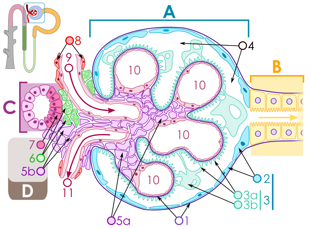
The key for the renal corpuscle figure is: A – [[Renal corpuscle]], B – [[Proximal tubule]], C – [[Distal convoluted tubule]], D – [[Juxtaglomerular apparatus]], 1. [[Basement membrane]] (Basal lamina), 2. [[Bowman's capsule]] – parietal layer, 3. [[Bowman's capsule]] – visceral layer, 3a. Pedicels (Foot processes from [[podocytes]]), 3b. [[Podocyte]], 4. [[Bowman's space]] (urinary space), 5a. [[Mesangium]] – Intraglomerular cell, 5b. [[Mesangium]] – Extraglomerular cell, 6. Granular cells ([[Juxtaglomerular cells]]), 7. [[Macula densa]], 8. [[Myocytes]] ([[smooth muscle]]), 9. [[Afferent arteriole]], 10. Glomerulus [[Capillaries]], 11. [[Efferent arteriole]]. | The key for the renal corpuscle figure is: A – [[Renal corpuscle]], B – [[Proximal tubule]], C – [[Distal convoluted tubule]], D – [[Juxtaglomerular apparatus]], 1. [[Basement membrane]] (Basal lamina), 2. [[Bowman's capsule]] – parietal layer, 3. [[Bowman's capsule]] – visceral layer, 3a. Pedicels (Foot processes from [[podocytes]]), 3b. [[Podocyte]], 4. [[Bowman's space]] (urinary space), 5a. [[Mesangium]] – Intraglomerular cell, 5b. [[Mesangium]] – Extraglomerular cell, 6. Granular cells ([[Juxtaglomerular cells]]), 7. [[Macula densa]], 8. [[Myocytes]] ([[smooth muscle]]), 9. [[Afferent arteriole]], 10. Glomerulus [[Capillaries]], 11. [[Efferent arteriole]]. | ||
Rapidly progressive glomerulonephritis is a disease of the kidney in which the renal function deteriorates very quickly in a matter of days. | |||
Atleast 50% reduction in GFR occurs in RPGN in a few days to weeks. | |||
RPGN occurs from severe and fast damage to the GBM which results in crescent formation,the main pathological finding in RPGN. | |||
=== Pathogenesis === | === Pathogenesis === | ||
* | * RPGN is characterized by severe and fast damage to the GBM that results in atleast 50% reduction in GFR in a few days. | ||
* | * The injury to GBM can be caused by multiple factors. | ||
* | * Crescent formation is the major pathological finding. | ||
** | * In some cases crescents might be absent. | ||
*** | |||
*** | ====== Cresent formation ====== | ||
*** | * Crescents are defined as 2 or more layers of proliferating cells in the Bowman's space. | ||
** | * It is a response that occurs following severe damage to the glomerulus. | ||
*** | * Crescents are formed when fibrin deposition occurs in Bowmans 'space. | ||
*** | * Fibrin formation in Bowman's space is a complex process which involves multiple components. | ||
*** | * Fibrin formation is precipitated by leakage of multiple cells and inflammation mediators through glomerular capliiary wall,GBM and Bowman's capsule. | ||
* | * This leakage occurs from damage caused by multiple factors: | ||
* | ** Anti GBM antiboides | ||
** Immune complexes | |||
** ANCA or vasculitis | |||
* The cells include coagulation proteins,macrophages,T cells and other inflammatory mediators. | |||
* This leads to fibrous tissue formation in the Bowman's space known as crescents. | |||
====== Glomerular injury ====== | |||
* Injury to the glomerulus is the initiating factor for crescent formation. | |||
* Injury can occur by the following | |||
*# Anti GBM antibodies-Type I RPGN | |||
*##** These are autoantibodies that cross react with type IV collagen of the GBM. | |||
*##** These can be produced due to genetic causes such as in Goodpasture diseases or they can be produced after viral URTI or cigarette smoking. | |||
*##** These autoantibodies react with the GBM resulting in IgG deposition over the GBM. | |||
*##** The IgG activates helper T cells that attract the inflammatory mediators to the GBM damaging the glomeruli. | |||
*##** This damage causes leakage of cells and inflammatory mediators resulting in crescent formation. | |||
*##** The anti GBM antibodies can affect the lungs as well as in Goodpasture syndrome resulting in glomerular necrosis and alveolar haemorrhages. | |||
2. Immune complex- Type II RPGN | |||
* Immune complexes are formed in certain infections, connective tissue diseases, side effects of some drugs and in some myeloproliferative disorders. | |||
* These immune complexes are deposited over the GBM. | |||
* The immune complexes activate the complement system which sets off the inflammatory process. | |||
* The complement cascade is activated, attracting inflammatory cells and mediators to the GBM. | |||
* The serum levels of c3 and c4 fall down and is an indicator of immune complex mediated glomerular injury. | |||
* This damages the glomeruli and causes leakage of cells and inflammatory mediators resulting in crescent formation. | |||
3. Pauci immune RPGN-Type III RPGN | |||
* No circulating immune complexes or antibodies. | |||
* Glomerular damage is caused by circulating ANCAs(anti nuclear cytoplasmic antibodies) . | |||
* ANCAs cause glomerular damage by releasing lytic enzymes from white blood cells such as neutrophils. | |||
* These lytic enzymes damage the GBM and cause leakage of circulating cells that result in fibrin formation in the Bowmans space. | |||
* ANCAs are associated with systemic vasculitis. | |||
== Microscopic pathology == | == Microscopic pathology == | ||
Revision as of 21:27, 18 July 2018
|
Rapidly progressive glomerulonephritis Microchapters |
|
Differentiating Rapidly progressive glomerulonephritis from other Diseases |
|---|
|
Diagnosis |
|
Treatment |
|
Case Studies |
|
Rapidly progressive glomerulonephritis pathophysiology On the Web |
|
American Roentgen Ray Society Images of Rapidly progressive glomerulonephritis pathophysiology |
|
FDA on Rapidly progressive glomerulonephritis pathophysiology |
|
CDC on Rapidly progressive glomerulonephritis pathophysiology |
|
Rapidly progressive glomerulonephritis pathophysiology in the news |
|
Blogs on Rapidly progressive glomerulonephritis pathophysiology |
|
Directions to Hospitals Treating Rapidly progressive glomerulonephritis |
|
Risk calculators and risk factors for Rapidly progressive glomerulonephritis pathophysiology |
Editor-In-Chief: C. Michael Gibson, M.S., M.D. [1]
Overview
Pathophysiology
Anatomy


The key for the renal corpuscle figure is: A – Renal corpuscle, B – Proximal tubule, C – Distal convoluted tubule, D – Juxtaglomerular apparatus, 1. Basement membrane (Basal lamina), 2. Bowman's capsule – parietal layer, 3. Bowman's capsule – visceral layer, 3a. Pedicels (Foot processes from podocytes), 3b. Podocyte, 4. Bowman's space (urinary space), 5a. Mesangium – Intraglomerular cell, 5b. Mesangium – Extraglomerular cell, 6. Granular cells (Juxtaglomerular cells), 7. Macula densa, 8. Myocytes (smooth muscle), 9. Afferent arteriole, 10. Glomerulus Capillaries, 11. Efferent arteriole.
Rapidly progressive glomerulonephritis is a disease of the kidney in which the renal function deteriorates very quickly in a matter of days.
Atleast 50% reduction in GFR occurs in RPGN in a few days to weeks.
RPGN occurs from severe and fast damage to the GBM which results in crescent formation,the main pathological finding in RPGN.
Pathogenesis
- RPGN is characterized by severe and fast damage to the GBM that results in atleast 50% reduction in GFR in a few days.
- The injury to GBM can be caused by multiple factors.
- Crescent formation is the major pathological finding.
- In some cases crescents might be absent.
Cresent formation
- Crescents are defined as 2 or more layers of proliferating cells in the Bowman's space.
- It is a response that occurs following severe damage to the glomerulus.
- Crescents are formed when fibrin deposition occurs in Bowmans 'space.
- Fibrin formation in Bowman's space is a complex process which involves multiple components.
- Fibrin formation is precipitated by leakage of multiple cells and inflammation mediators through glomerular capliiary wall,GBM and Bowman's capsule.
- This leakage occurs from damage caused by multiple factors:
- Anti GBM antiboides
- Immune complexes
- ANCA or vasculitis
- The cells include coagulation proteins,macrophages,T cells and other inflammatory mediators.
- This leads to fibrous tissue formation in the Bowman's space known as crescents.
Glomerular injury
- Injury to the glomerulus is the initiating factor for crescent formation.
- Injury can occur by the following
- Anti GBM antibodies-Type I RPGN
- These are autoantibodies that cross react with type IV collagen of the GBM.
- These can be produced due to genetic causes such as in Goodpasture diseases or they can be produced after viral URTI or cigarette smoking.
- These autoantibodies react with the GBM resulting in IgG deposition over the GBM.
- The IgG activates helper T cells that attract the inflammatory mediators to the GBM damaging the glomeruli.
- This damage causes leakage of cells and inflammatory mediators resulting in crescent formation.
- The anti GBM antibodies can affect the lungs as well as in Goodpasture syndrome resulting in glomerular necrosis and alveolar haemorrhages.
- Anti GBM antibodies-Type I RPGN
2. Immune complex- Type II RPGN
- Immune complexes are formed in certain infections, connective tissue diseases, side effects of some drugs and in some myeloproliferative disorders.
- These immune complexes are deposited over the GBM.
- The immune complexes activate the complement system which sets off the inflammatory process.
- The complement cascade is activated, attracting inflammatory cells and mediators to the GBM.
- The serum levels of c3 and c4 fall down and is an indicator of immune complex mediated glomerular injury.
- This damages the glomeruli and causes leakage of cells and inflammatory mediators resulting in crescent formation.
3. Pauci immune RPGN-Type III RPGN
- No circulating immune complexes or antibodies.
- Glomerular damage is caused by circulating ANCAs(anti nuclear cytoplasmic antibodies) .
- ANCAs cause glomerular damage by releasing lytic enzymes from white blood cells such as neutrophils.
- These lytic enzymes damage the GBM and cause leakage of circulating cells that result in fibrin formation in the Bowmans space.
- ANCAs are associated with systemic vasculitis.
Microscopic pathology
{{#ev:youtube|CqSyj4cVZPE}}