Pulmonary hypertension chest x ray: Difference between revisions
Jump to navigation
Jump to search
No edit summary |
No edit summary |
||
| Line 11: | Line 11: | ||
===Findings on Chest x-ray=== | ===Findings on Chest x-ray=== | ||
# Hilar pulmonary arterial dilation. | # [[Hilar]] pulmonary arterial dilation. | ||
# Loss of peripheral blood vessel markings. | # Loss of peripheral blood vessel markings. | ||
# Enlarged right atrium and right ventricle in advanced diseases. | # Enlarged right atrium and right ventricle in advanced diseases. | ||
| Line 18: | Line 18: | ||
<gallery> | <gallery> | ||
Image:336139-361242-1175.jpg|This is a posteroanterior radiograph revealing enlarged pulmonary arteries in a patient with Atrial septal defect. | Image:336139-361242-1175.jpg|This is a posteroanterior radiograph revealing enlarged pulmonary arteries in a patient with [[Atrial septal defect]]. | ||
Image:Pulmon4.gif | Image:Pulmon4.gif | ||
Image:CXR for a patient with advanced Histiocytosis X associated with severe pulmonary hypertension.jpeg|Chest x-ray for a patient with advanced Histiocytosis X associated with severe pulmonary hypertension | Image:CXR for a patient with advanced [[Histiocytosis X]] associated with severe pulmonary hypertension.jpeg|Chest x-ray for a patient with advanced Histiocytosis X associated with severe pulmonary hypertension | ||
</gallery> | </gallery> | ||
Revision as of 17:55, 25 September 2012
|
Pulmonary Hypertension Microchapters |
|
Diagnosis |
|---|
|
Treatment |
|
Case Studies |
|
Pulmonary hypertension chest x ray On the Web |
|
American Roentgen Ray Society Images of Pulmonary hypertension chest x ray |
|
Risk calculators and risk factors for Pulmonary hypertension chest x ray |
Editor-In-Chief: C. Michael Gibson, M.S., M.D. [1], Richard Channick, M.D.; Assistant Editor(s)-in-Chief: Ralph Matar.
Overview
Chest X Ray
Chest x ray in a patient with pulmonary hypertension
- Chest x-ray is abnormal in 90% of patients with pulmonary hypertension at the time of diagnosis. However, no correlation have been found between the degree of severity of pulmonary hypertension and the findings on chest x-rays.
- It allows exclusion of moderate to severe lung diseases and pulmonary venous hypertension due to left heart disease.
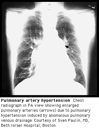
Findings on Chest x-ray
- Hilar pulmonary arterial dilation.
- Loss of peripheral blood vessel markings.
- Enlarged right atrium and right ventricle in advanced diseases.
Typical chest x-rays
-
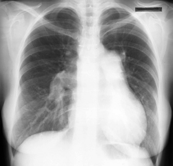
This is a posteroanterior radiograph revealing enlarged pulmonary arteries in a patient with Atrial septal defect.
-