Phyllodes tumor
|
WikiDoc Resources for Phyllodes tumor |
|
Articles |
|---|
|
Most recent articles on Phyllodes tumor Most cited articles on Phyllodes tumor |
|
Media |
|
Powerpoint slides on Phyllodes tumor |
|
Evidence Based Medicine |
|
Clinical Trials |
|
Ongoing Trials on Phyllodes tumor at Clinical Trials.gov Trial results on Phyllodes tumor Clinical Trials on Phyllodes tumor at Google
|
|
Guidelines / Policies / Govt |
|
US National Guidelines Clearinghouse on Phyllodes tumor NICE Guidance on Phyllodes tumor
|
|
Books |
|
News |
|
Commentary |
|
Definitions |
|
Patient Resources / Community |
|
Patient resources on Phyllodes tumor Discussion groups on Phyllodes tumor Patient Handouts on Phyllodes tumor Directions to Hospitals Treating Phyllodes tumor Risk calculators and risk factors for Phyllodes tumor
|
|
Healthcare Provider Resources |
|
Causes & Risk Factors for Phyllodes tumor |
|
Continuing Medical Education (CME) |
|
International |
|
|
|
Business |
|
Experimental / Informatics |
Editor-In-Chief: C. Michael Gibson, M.S., M.D. [1] Associate Editor(s)-in-Chief: Maria Fernanda Villarreal, M.D. [2]
Synonyms and keywords: Cystosarcoma phyllodes; Cystosarcoma phylloides; Phyllodes tumours
Overview
Phyllodes tumor (also known as cystosarcoma phyllodes), is a typically large, fast-growing mass that arises from the periductal stromal cells of the breast. Phyllodes tumors account for less than 1% of all breast neoplasms. Phyllodes tumor was first discovered by Johannes Muller, a German physician in 1838. Phyllodes tumor may be classified according to histological grade into 3 subtypes: benign, borderline, and malignant. The pathogenesis of phyllodes tumor is characterized by the overgrowth of stromal cells. Genes involved in the development of phyllodes tumor, include: p53 gene, EGFR gene, and IGF1R gene. There are no established causes for phyllodes tumor. Phyllodes tumors are rare, and approximately 6% of all phyllodes tumors are malignant. The diagnosis of phyllodes tumor is preferably made with core needle biopsy. The majority of patients with phyllodes tumor remain asymptomatic for years. The most important early clinical feature is a rapid growing palpable mass. Mammography is the imaging modality of choice for phyllodes tumor, characteristic findings include: large rounded oval or lobulated mass, well circumscribed, and smooth margins. Wide local excision is the most common approach to the treatment of phyllodes tumor.[1]
Historical Perspective
- Phyllodes tumor was first discovered by Johannes Muller, a German physician in 1838.[2]
Classification
- Phyllodes tumor may be classified according to histological grade into 3 subtypes:[2]
- Benign (most common)
- Borderline
- Malignant
Pathophysiology
- The pathogenesis of phyllodes tumor is characterized by the overgrowth of stromal cells.[1]
- Phyllodes tumor arises from the periductal stromal cells of the breast, which are normally involved in the supportive function of the parenchymal tissue.[1]
- Genes involved in the development of phyllodes tumor, include:[1]
- On gross pathology, characteristic findings of phyllodes tumor, include:
- Cleft/leaf-like structures
- Friable mass
- Well-defined margins
- On microscopic histopathological analysis,characteristic findings of phyllodes tumor, include:
- Large slit-like spaces
- Cellular myxoid stroma
- Infiltrative border
- Stromal overgrowth
Causes
- There are no established causes for phyllodes tumor.[2]
Differentiating Phyllodes Tumor from Other Diseases
- Phyllodes tumor must be differentiated from other diseases that cause a breast mass, such as:[2]
- Juvenile fibroadenoma (most common)
- Breast abscess
- Adenocarcinoma
- Mastitis
Epidemiology and Demographics
- The prevalence of phyllodes tumor is approximately 0.05 per 100,000 individuals worldwide.[1]
- Approximately 6% of all phyllodes tumors are malignant.
- Phyllodes tumor accounts for less than 0.3-1% of all breast neoplasms.[1]
Age
- Patients of all age groups may develop phyllodes tumor.
- However, phyllodes tumor is more commonly reported in adult women between 40 and 60 years.[2]
Gender
- Females are more commonly affected with phyllodes tumor than males.
Race
- Latina-whites are more commonly affected with phyllodes tumor.
Risk Factors
- There are no risk factors associated in the development of phyllodes tumor.[2]
- In some cases, patients with Li-Fraumeni syndrome may have an increased risk.
Natural History, Complications and Prognosis
- The majority of patients with phyllodes tumor remain asymptomatic for years.[1]
- The most important early clinical feature is a rapid growing palpable mass.
- If left untreated, patients with phyllodes tumor may progress to develop malignant degeneration.
- Complications of phyllodes tumor are usually related to surgery (mastectomy).[2]
- Common complications, include:[1]
- Infection
- Seroma formation
- Local or distant recurrence
- Prognosis is generally good, and the 5 year survival rate of patients with phyllodes tumor is approximately 60 to 80%.[2]
Diagnosis
Diagnostic Criteria
- The diagnosis of phyllodes tumor is preferably made with core needle biopsy.[1]
Symptoms
- Phyllodes tumor is usually asymptomatic.[2]
- There are no remarkable symptoms of phyllodes tumor.
- A directed history should be obtained to ascertain:
- Family history of breast malignancy
Physical Examination
- Patients with phyllodes tumor are usually well-appearing.
- Breast physical examination may be remarkable for:
- Breast mass, with the following characteristics:[2]
- Firm
- Mobile
- Well-circumscribed
- Non-tender
Laboratory Findings
- There are no specific laboratory findings associated with phyllodes tumor.
Imaging Findings
- Mammography is the imaging modality of choice for phyllodes tumor.[1]
- On mammography, findings of phyllodes tumor include:[2]
- Non specific large rounded oval or lobulated mass
- Generally well circumscribed
- Lesions with smooth margins
- A radiolucent halo may be present
- Calcification (typically coarse and plaque like) may be seen in a very small proportion.
-
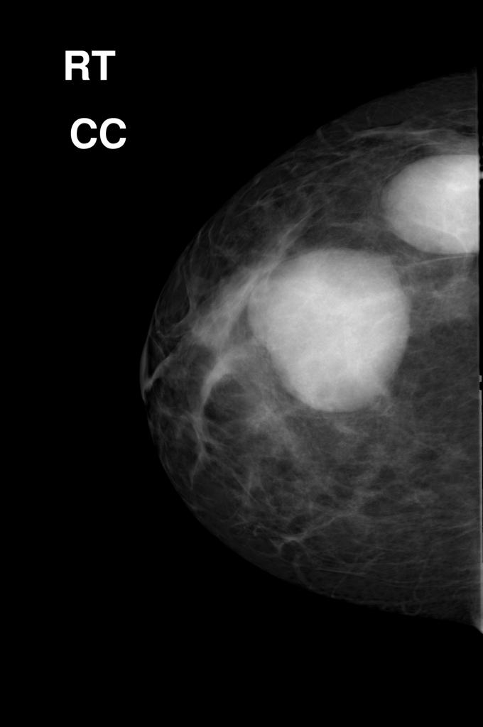
Mammography of phyllodes tumor
Adapted from Radiopedia

- On ultrasound, findings of phyllodes tumor include:[1]
- Inhomogeneous
- Solid-appearing mass
- Solid mass containing single or multiple, round or cleft like cystic spaces
- Posterior acoustic enhancement strongly suggests the diagnosis of phyllodes tumor
- Vascularisation is usually present in the solid components
- On MRI, findings of phyllodes tumor include:
- T1: usually of low signal
- T2: can be variable ranging from homogenous low 8 to high 4-5 signal
- T1 C+ (Gd): the solid components enhance after contrast administration
- Dynamic contrast: the kinetic curve pattern can be gradual slow or have rapid enhancement
Treatment
Medical Therapy
- There is no medical therapy for phyllodes tumor.
- Existing medical therapies, such as hormonal therapy and chemotherapy are proven to be ineffective among patients with phyllodes tumor.
Surgery
- Surgery is the mainstay of therapy for phyllodes tumor.[1]
- Wide local excision is the most common approach to the treatment of phyllodes tumor.
- The risk of developing local recurrence or metastases among patients with phyllodes tumor is related to the histologic grade.
Prevention
- Effective measures for the secondary prevention of phyllodes tumor include periodical self-breast examination, and routine mammography.
- Once diagnosed and successfully treated, patients with phyllodes tumor are followed-up periodically.
- Follow-up testing, includes: ultrasound examination, breast exam, and mammography.
References
- ↑ 1.00 1.01 1.02 1.03 1.04 1.05 1.06 1.07 1.08 1.09 1.10 1.11 Phyllodes tumor. Dr Henry Knipe. Radiopedia. http://radiopaedia.org/articles/phyllodes-tumour Accessed on May 19, 2016
- ↑ 2.00 2.01 2.02 2.03 2.04 2.05 2.06 2.07 2.08 2.09 2.10 Phyllodes tumor. Wikipedia. https://en.wikipedia.org/wiki/Phyllodes_tumor