Oral candidiasis physical examination
|
Oral candidiasis Microchapters |
|
Diagnosis |
|---|
|
Treatment |
|
Case Studies |
|
Oral candidiasis physical examination On the Web |
|
American Roentgen Ray Society Images of Oral candidiasis physical examination |
|
Risk calculators and risk factors for Oral candidiasis physical examination |
Editor-In-Chief: C. Michael Gibson, M.S., M.D. [1]
Physical Examination
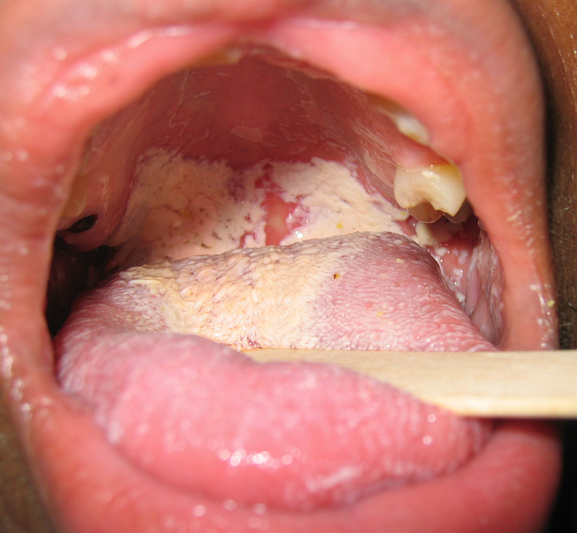
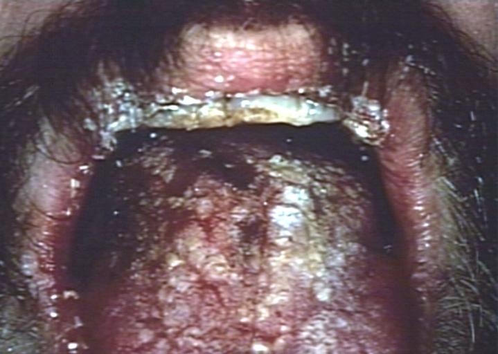
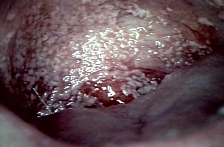
Oral infections of candidia usually appear as thick white or cream-colour deposits. Underlying the deposits the mucosa of the mouth may appear inflamed (red and possibly slightly raised). Oral lesions are painless, white patches in the mouth.
-
-
Oral manifestations of HIV infection and AIDS. Chronic oral candidiasis in patient with AIDS. Image courtesy of Professor Peter Anderson DVM PhD and published with permission. © PEIR, University of Alabama at Birmingham, Department of Pathology
-
Soft palate showing extensive oral candidiasis in patient with AIDS. Image courtesy of Professor Peter Anderson DVM PhD and published with permission. © PEIR, University of Alabama at Birmingham, Department of Pathology


