Multiple sclerosis MRI
|
Multiple sclerosis Microchapters |
|
Diagnosis |
|---|
|
Treatment |
|
Case Studies |
|
Multiple sclerosis MRI On the Web |
|
American Roentgen Ray Society Images of Multiple sclerosis MRI |
|
Risk calculators and risk factors for Multiple sclerosis MRI |
Editor-In-Chief: C. Michael Gibson, M.S., M.D. [1]
Overview
MRI
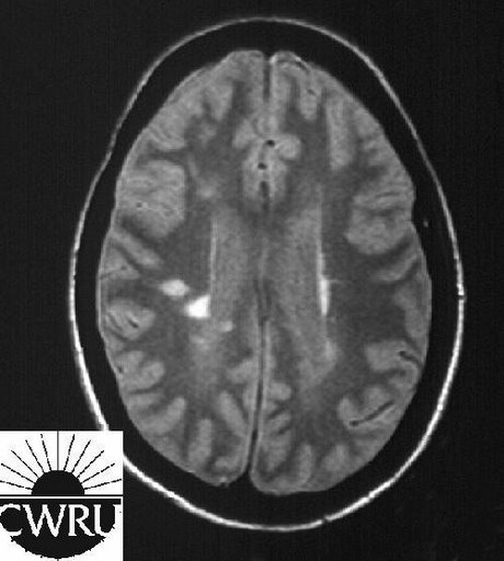
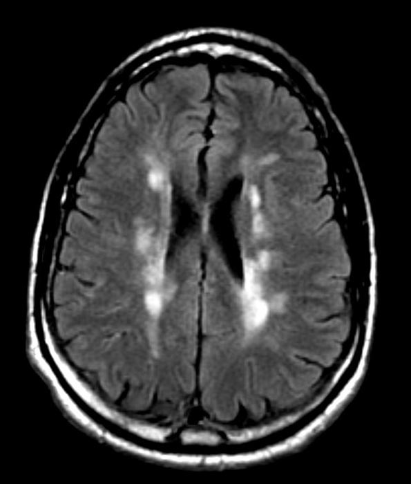
On MRI, multiple sclerosis is characterized by cerebral and/or spinal cord plaques which are demyelinating areas.[1] These lesions are commonly ovoid, and located in periventricular white matter, cerebellum and brain stem.[2]
-
T1-weighted MRI scans (post-contrast) of same brain slice at monthly intervals. Bright spots indicate active lesions.
-
Multiple Sclerosis
Patient #1
-
Multiple sclerosis
-
Multiple sclerosis
-
Multiple sclerosis
-
Multiple sclerosis
-
Multiple sclerosis
-
Multiple sclerosis
Patient #2: Contrast enchancement of several lesions indicates active disease
-
-
-
-
GAD enhanced T1
References
- ↑ Trapp BD, Peterson J, Ransohoff RM, Rudick R, Mörk S, Bö L (January 1998). "Axonal transection in the lesions of multiple sclerosis". N. Engl. J. Med. 338 (5): 278–85. doi:10.1056/NEJM199801293380502. PMID 9445407.
- ↑ Fazekas F, Barkhof F, Filippi M, Grossman RI, Li DK, McDonald WI, McFarland HF, Paty DW, Simon JH, Wolinsky JS, Miller DH (August 1999). "The contribution of magnetic resonance imaging to the diagnosis of multiple sclerosis". Neurology. 53 (3): 448–56. PMID 10449103.


