Multiple sclerosis MRI: Difference between revisions
No edit summary |
No edit summary |
||
| Line 6: | Line 6: | ||
==MRI== | ==MRI== | ||
The core of diagnosing [[MS]] is to show disseminating [[lesions]] in space and time. Before [[MRI]] becomes a common imaging technique for [[diagnosis]] [[MS]], clinical presentation was the only tool. Nowadays new [[diagnostic criteria]] ( | The core of diagnosing [[MS]] is to show disseminating [[lesions]] in space and time. Before [[MRI]] becomes a common imaging technique for [[diagnosis]] [[MS]], clinical presentation was the only tool. Nowadays new [[diagnostic criteria]] ([[mcDonald criteria]]) are focusing on [[MRI]] finding in addition to clinical presentation of patients.<ref name="pmid11456302">{{cite journal |vauthors=McDonald WI, Compston A, Edan G, Goodkin D, Hartung HP, Lublin FD, McFarland HF, Paty DW, Polman CH, Reingold SC, Sandberg-Wollheim M, Sibley W, Thompson A, van den Noort S, Weinshenker BY, Wolinsky JS |title=Recommended diagnostic criteria for multiple sclerosis: guidelines from the International Panel on the diagnosis of multiple sclerosis |journal=Ann. Neurol. |volume=50 |issue=1 |pages=121–7 |date=July 2001 |pmid=11456302 |doi= |url=}}</ref> On [[MRI]], multiple sclerosis is characterized by [[cerebral]] and/or [[spinal cord]] plaques which are [[demyelinating]] areas.<ref name="pmid9445407">{{cite journal |vauthors=Trapp BD, Peterson J, Ransohoff RM, Rudick R, Mörk S, Bö L |title=Axonal transection in the lesions of multiple sclerosis |journal=N. Engl. J. Med. |volume=338 |issue=5 |pages=278–85 |date=January 1998 |pmid=9445407 |doi=10.1056/NEJM199801293380502 |url=}}</ref> | ||
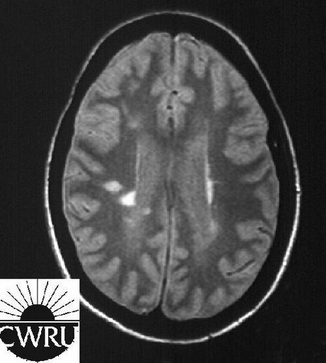
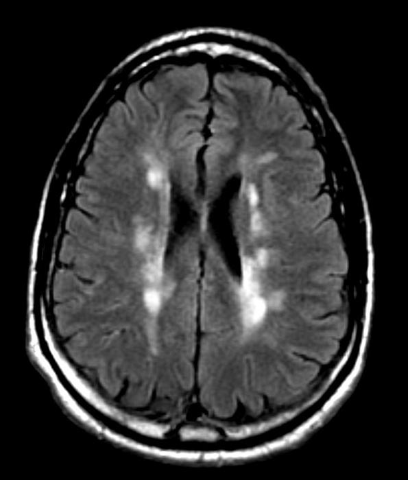
These [[Lesion|lesions]] are commonly ovoid, and located in periventricular [[white matter]], [[cerebellum]] and [[brain stem]].<ref name="pmid10449103">{{cite journal |vauthors=Fazekas F, Barkhof F, Filippi M, Grossman RI, Li DK, McDonald WI, McFarland HF, Paty DW, Simon JH, Wolinsky JS, Miller DH |title=The contribution of magnetic resonance imaging to the diagnosis of multiple sclerosis |journal=Neurology |volume=53 |issue=3 |pages=448–56 |date=August 1999 |pmid=10449103 |doi= |url=}}</ref> These lesions are hyperintense on T2 sections of [[MRI]]. There are also [[gray matter]] lesions, presents as hypointensity in T2-weighted and are correlated with patients physical [[disability]] and brain [[atrophy]].<ref name="pmid10683822">{{cite journal |vauthors=Bakshi R, Shaikh ZA, Janardhan V |title=MRI T2 shortening ('black T2') in multiple sclerosis: frequency, location, and clinical correlation |journal=Neuroreport |volume=11 |issue=1 |pages=15–21 |date=January 2000 |pmid=10683822 |doi= |url=}}</ref><ref name="pmid11266686">{{cite journal |vauthors=Bakshi R, Dmochowski J, Shaikh ZA, Jacobs L |title=Gray matter T2 hypointensity is related to plaques and atrophy in the brains of multiple sclerosis patients |journal=J. Neurol. Sci. |volume=185 |issue=1 |pages=19–26 |date=March 2001 |pmid=11266686 |doi= |url=}}</ref> | These [[Lesion|lesions]] are commonly ovoid, and located in periventricular [[white matter]], [[cerebellum]] and [[brain stem]].<ref name="pmid10449103">{{cite journal |vauthors=Fazekas F, Barkhof F, Filippi M, Grossman RI, Li DK, McDonald WI, McFarland HF, Paty DW, Simon JH, Wolinsky JS, Miller DH |title=The contribution of magnetic resonance imaging to the diagnosis of multiple sclerosis |journal=Neurology |volume=53 |issue=3 |pages=448–56 |date=August 1999 |pmid=10449103 |doi= |url=}}</ref> These lesions are hyperintense on T2 sections of [[MRI]]. There are also [[gray matter]] lesions, presents as hypointensity in T2-weighted and are correlated with patients physical [[disability]] and brain [[atrophy]].<ref name="pmid10683822">{{cite journal |vauthors=Bakshi R, Shaikh ZA, Janardhan V |title=MRI T2 shortening ('black T2') in multiple sclerosis: frequency, location, and clinical correlation |journal=Neuroreport |volume=11 |issue=1 |pages=15–21 |date=January 2000 |pmid=10683822 |doi= |url=}}</ref><ref name="pmid11266686">{{cite journal |vauthors=Bakshi R, Dmochowski J, Shaikh ZA, Jacobs L |title=Gray matter T2 hypointensity is related to plaques and atrophy in the brains of multiple sclerosis patients |journal=J. Neurol. Sci. |volume=185 |issue=1 |pages=19–26 |date=March 2001 |pmid=11266686 |doi= |url=}}</ref> | ||
Revision as of 06:30, 1 March 2018
|
Multiple sclerosis Microchapters |
|
Diagnosis |
|---|
|
Treatment |
|
Case Studies |
|
Multiple sclerosis MRI On the Web |
|
American Roentgen Ray Society Images of Multiple sclerosis MRI |
|
Risk calculators and risk factors for Multiple sclerosis MRI |
Editor-In-Chief: C. Michael Gibson, M.S., M.D. [1]
Overview
MRI
The core of diagnosing MS is to show disseminating lesions in space and time. Before MRI becomes a common imaging technique for diagnosis MS, clinical presentation was the only tool. Nowadays new diagnostic criteria (mcDonald criteria) are focusing on MRI finding in addition to clinical presentation of patients.[1] On MRI, multiple sclerosis is characterized by cerebral and/or spinal cord plaques which are demyelinating areas.[2] These lesions are commonly ovoid, and located in periventricular white matter, cerebellum and brain stem.[3] These lesions are hyperintense on T2 sections of MRI. There are also gray matter lesions, presents as hypointensity in T2-weighted and are correlated with patients physical disability and brain atrophy.[4][5]
-

T1-weighted MRI scans (post-contrast) of same brain slice at monthly intervals. Bright spots indicate active lesions.
-
Multiple Sclerosis


Patient #1
-

Multiple sclerosis
-

Multiple sclerosis
-

Multiple sclerosis
-

Multiple sclerosis
-

Multiple sclerosis
-

Multiple sclerosis


Patient #2: Contrast enchancement of several lesions indicates active disease
-

-
-

-

GAD enhanced T1


References
- ↑ McDonald WI, Compston A, Edan G, Goodkin D, Hartung HP, Lublin FD, McFarland HF, Paty DW, Polman CH, Reingold SC, Sandberg-Wollheim M, Sibley W, Thompson A, van den Noort S, Weinshenker BY, Wolinsky JS (July 2001). "Recommended diagnostic criteria for multiple sclerosis: guidelines from the International Panel on the diagnosis of multiple sclerosis". Ann. Neurol. 50 (1): 121–7. PMID 11456302.
- ↑ Trapp BD, Peterson J, Ransohoff RM, Rudick R, Mörk S, Bö L (January 1998). "Axonal transection in the lesions of multiple sclerosis". N. Engl. J. Med. 338 (5): 278–85. doi:10.1056/NEJM199801293380502. PMID 9445407.
- ↑ Fazekas F, Barkhof F, Filippi M, Grossman RI, Li DK, McDonald WI, McFarland HF, Paty DW, Simon JH, Wolinsky JS, Miller DH (August 1999). "The contribution of magnetic resonance imaging to the diagnosis of multiple sclerosis". Neurology. 53 (3): 448–56. PMID 10449103.
- ↑ Bakshi R, Shaikh ZA, Janardhan V (January 2000). "MRI T2 shortening ('black T2') in multiple sclerosis: frequency, location, and clinical correlation". Neuroreport. 11 (1): 15–21. PMID 10683822.
- ↑ Bakshi R, Dmochowski J, Shaikh ZA, Jacobs L (March 2001). "Gray matter T2 hypointensity is related to plaques and atrophy in the brains of multiple sclerosis patients". J. Neurol. Sci. 185 (1): 19–26. PMID 11266686.