Melanoma surgery
Editor-In-Chief: C. Michael Gibson, M.S., M.D. [1]
|
Melanoma Microchapters |
|
Diagnosis |
|---|
|
Treatment |
|
Case Studies |
|
Melanoma surgery On the Web |
|
American Roentgen Ray Society Images of Melanoma surgery |
Overview
Minor surgery
Moles that are irregular in color or shape are suspicious of a malignant or a premalignant melanoma. Following a visual examination and a dermatoscopic exam (an instrument that illuminates a mole, revealing its underlying pigment and vascular network structure), the doctor may biopsy the suspicious mole. If it is malignant, the mole and an area around it needs excision. This will require a referral to a surgeon or dermatologist.
The diagnosis of melanoma requires experience, as early stages may look identical to harmless moles or not have any color at all. Where any doubt exists, the patient will be referred to a specialist dermatologist. Beyond this expert knowledge a biopsy performed under local anesthesia is often required to assist in making or confirming the diagnosis and in defining the severity of the melanoma.
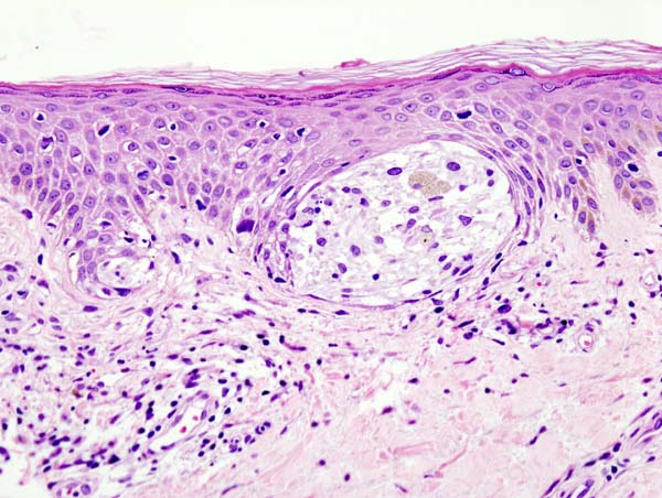
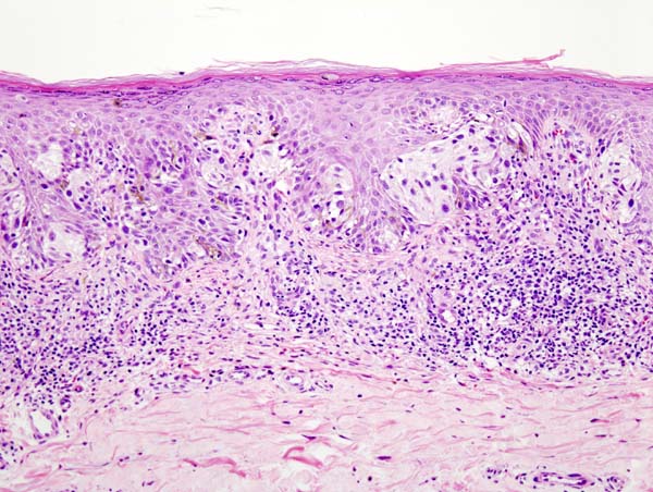
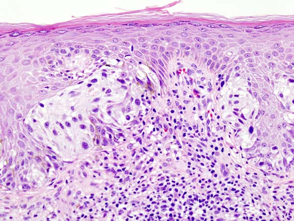
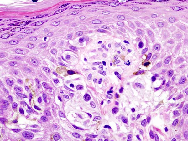
Excisional biopsy is the management of choice; this is where the suspect lesion is totally removed with an adequate ellipse of surrounding skin and tissue.[1] The biopsy will include the epidermal, dermal, and subcutaneous layers of the skin, enabling the histopathologist to determine the depth of penetration of the melanoma by microscopic examination. This is described by Clark's level (involvement of skin structures) and Breslow's depth (measured in millimeters).
If an excisional biopsy is not possible in certain larger pigmented lesions, a punch biopsy may be performed by a specialist hospital doctor, using a surgical punch (an instrument similar to a tiny cookie cutter with a handle, with an opening ranging in size from 1 to 6 mm). The punch is used to remove a plug of skin (down to the subcutaneous layer) from a portion of a large suspicious lesion, for histopathological examination.
References
- ↑ Swanson N, Lee K, Gorman A, Lee H (2002). "Biopsy techniques. Diagnosis of melanoma". Dermatol Clin. 20 (4): 677–80. PMID 12380054.


