Melanoma overview
Editor-In-Chief: C. Michael Gibson, M.S., M.D. [2]
|
Melanoma Microchapters |
|
Diagnosis |
|---|
|
Treatment |
|
Case Studies |
|
Melanoma overview On the Web |
|
American Roentgen Ray Society Images of Melanoma overview |
Overview
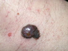
Melanoma is a malignant tumor of melanocytes which are found predominantly in skin but also in the bowel and the eye (see uveal melanoma). It is one of the rarer types of skin cancer but causes the majority of skin cancer related deaths.[1][2] Despite many years of intensive laboratory and clinical research, the sole effective cure is surgical resection of the primary tumor before it achieves a thickness greater than 1 mm.
Around 160,000 new cases of melanoma are diagnosed worldwide each year, and it is more frequent in males and caucasians.[3] It is more common in caucasian populations living in sunny climates than other groups.[4] According to the WHO Report about 48,000 melanoma related deaths occur worldwide per annum.[5]
The treatment includes surgical removal of the tumor; adjuvant treatment; chemo- and immunotherapy, or radiation therapy.
Melanomas also occur in horses, see equine melanoma.
Sometimes the skin lesion may bleed, itch, or ulcerate, although this is a very late sign. A slow-healing lesion should be watched closely, as that may be a sign of melanoma. Be aware also that in circumstances that are still poorly understood, melanomas may "regress" or spontaneously become smaller or invisible - however the malignancy is still present. Amelanotic (colorless or flesh-colored) melanomas do not have pigment and may not even be visible. Lentigo maligna, a superficial melanoma confined to the topmost layers of the skin (found primarily in older patients) is often described as a "stain" on the skin. Some patients with metastatic melanoma do not have an obvious detectable primary tumor.
Types of primary melanoma

The most common types of Melanoma in the skin:
- superficial spreading melanoma (SSM)
- nodular melanoma
- acral lentiginous melanoma
- lentigo maligna (melanoma)
Any of the above types may produce melanin (and be dark in colour) or not (and be amelanotic - not dark). Similarly any subtype may show desmoplasia (dense fibrous reaction with neurotropism) which is a marker of aggressive behaviour and a tendency to local recurrence.
Elsewhere:
- clear cell sarcoma (Melanoma of Soft Parts)
- mucosal melanoma
- uveal melanoma
Equine melanoma
Melanomas are also not uncommon in horses, being largely confined to grey (or white) animals - 80% of such pale horses will develop melanomata by 15 years of age[6]; of these, 66% are slow growing but all may be classified as malignant[6]. Surgical excision may be attempted in some cases, if the tumours are limited in extent and number. However, they are often multiple (especially in older animals) and perineal tumours are notoriously difficult to excise. Often, a position of "benign neglect" is assumed, especially if the tumours are not causing any clinical problems. Medical therapy with cimetidine (2.5-4.0mg/kg three times daily for 2 months or more)[7] is also an option, although it has a lower success rate than surgery and cryosurgery[8].
References
- ↑ Melanoma Death Rate Still Climbing
- ↑ Cancer Stat Fact Sheets
- ↑ Ries LAG, et al, eds. SEER Cancer Statistics Review, 1975-2000. Bethesda, MD: National Cancer Institute; 2003: Tables XVI-1-9.
- ↑ Parkin D, Bray F, Ferlay J, Pisani P. "Global cancer statistics, 2002". CA Cancer J Clin. 55 (2): 74–108. PMID 15761078.Full text
- ↑ Lucas, R. Global Burden of Disease of Solar Ultraviolet Radiation, Environmental Burden of Disease Series, July 25, 2006; No. 13. News release, World Health Organization
- ↑ 6.0 6.1 Centre for Comparitive Oncology [1], accessed at 2220 on 12th July
- ↑ Warnick, LD, Graham, ME, and Valentine, BA (1995) "Evaluation of cimetidine treatment for melanomas in seven horses" Equine Practice, 17(7): 16-22, 1995
- ↑ RJ Rose & DR Hodson, Manual of Equine Practice (p. 498) 2000