Hymenolepiasis
Causes, incidence, and risk factors

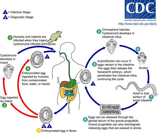
Hymenolepis worms live in warm climates and are common in the southern USA. The eggs of these worms are ingested by insects, and mature into a life form referred to as a "cysticercoid" in the insect. In H. nana, the insect is always a beetle. Humans and other animals become infected when they intentionally or unintentionally eat material contaminated by insects. In an infected person, it is possible for the worm's entire life-cycle to be completed in the bowel, so infection can persist for years. Hymenolepis nana infections are much more common than Hymenolepis diminuta infections in humans because, in addition to being spread by insects, the disease can be spread directly from person to person by eggs in feces. When this happens, H. nana oncosphere larvae encyst in the intestinal wall and develop into cysticercoids and then adults. These infections were previously common in the southeastern USA, and have been described in crowded environments and individuals confined to institutions. However, the disease occurs throughout the world. H. nana infections can grow worse over time because, unlike in most tapeworms, H. nana eggs can hatch and develop without ever leaving the definitive host.
A study in Connecticut found that one third of rats sold in pet stores were infected with H. nana and concluded that these and other rodents sold in pet stores pose a potential threat to public health.
Screening for activity against H. nana
H. nana in mice
Used because - Human infection—easily maintained in mice - Armed scolex similar to other pathogenic tapeworms - Corresponds to other tapeworms in its sensitivity to standard anthelmintics
Methods
  Mature worms collected from infected mice
  Terminal gravid proglottids removed, crushed under coverslip—eggs removed
Eggs containing hooklets (mature) counted 0.2 ml stock soln. containing 1000 eggs/ml given to each mouse. Adult worm develops- 15-17 days. Test drug given orally – autopsied on 3rd day Std. drug given Intestine examined under dissecting microscope for worms/ scolex Response – no. of mice cleared.
Symptoms
It is not clear that hymenolepiasis necessarily have any symptoms. The symptoms of hymenolepiasis are traditionally described as abdominal pain, loss of appetite (anorexia), itching around the anus, irritability and diarrhea. However, in one study of 25 patients conducted in Peru, successful treatment of the infection made no significant difference to symptoms.[1] Some authorities report that heavily infected cases are more likely to be symptomatic.[2][3]
Signs and tests
Examination of the stool for eggs and parasites confirms the diagnosis. The eggs and proglottids of H. nana are smaller than H. diminuta. Proglottids of both are relatively wide and have three testes. Identifying the parasites to the species level is often unnecessary from a medical perspective, as the treatment is the same for both.
Treatment
Praziquantel as a single dose (25 mg/kg) is the current treatment of choice for hymenolepiasis and has an efficacy of 96%. Single dose albendazole (400 mg) is also very efficacious (>95%). Niclosamide has also been used.
A three-day course of nitazoxanide is 75–93% efficacious. The dose is 1g daily for adults and children over 12; 400mg daily for children aged 4 to 11 years; and 200mg daily for children aged 3 years or younger.[1][4][5]
Prognosis
Cure rates are extremely good with modern treatments, but it is unclear that successful cure results in any symptomatic benefit to patients.[1]
Complications
- abdominal discomfort
- dehydration from prolonged diarrhea
Prevention
Good hygiene, public health and sanitation programs, and elimination of rats help prevent the spread of hymenolepiasis.
Source
- Hymenolepiasis. Medline Plus.
References
- ↑ 1.0 1.1 1.2 Chero JC, Saito M, Bustos JA; et al. (2007). "Hymenolepis nana infection: symptoms and response to nitazoxanide in field conditions". Trans R Soc Trop Med Hyg. 101 (2): 203&ndash, 5. doi:10.1016/j.trstmh.2006.04.004.
- ↑ Chitchang S, Plamjinda T, Yodmani B, Radomyos P. (1985). "Relationship between severity of the symptom and the number of Hymenolepis nana after treatment". J Med Assoc Thai. 68: 423&ndash, 26.
- ↑ Schantz PM. (1996). "Tapeworm (cestodiasis)". Gastroenterol Clin North Am. 25: 637&ndash, 53.
- ↑ Ortiz JJ, Favennec L, Chegne NL, Gargala G. (2002). "Comparative clinical studis of nitazoxanide, albendazole and praziquantel in the treatment of ascariasis, trichuriasis, and hymenolepiasis in children from Peru". Trans R Soc Trop med Hyg. 96: 193&ndash, 96. PMID 12055813.
- ↑ Reomero-Cabello R, Guerro LR, Munez-Gracia MR, Geyne Cruz A. (1997). "Nitazoxanide for the treatment of intestinal protozoan and helminthic infections in México". Trans R Soc Trop Med Hyg. 91: 701&ndash, 3.
Template:Helminthiases it:Hymenolepis nana Template:WikiDoc Sources