Hodgkin's lymphoma CT: Difference between revisions
No edit summary |
m (Bot: Removing from Primary care) |
||
| (14 intermediate revisions by 4 users not shown) | |||
| Line 1: | Line 1: | ||
__NOTOC__ | __NOTOC__ | ||
{{Hodgkin's lymphoma}} | {{Hodgkin's lymphoma}} | ||
{{CMG}}; {{AE}} {{AS}} | |||
==Overview== | |||
Chest, abdomen, and pelvis [[CT]] scan may be helpful in the diagnosis of Hodgkin's lymphoma. | |||
==CT== | |||
Chest, abdomen, and pelvis [[CT]] scan may be helpful in the diagnosis of Hodgkin's lymphoma. | |||
[[CT]] scan is used to:<ref>Hodgkin-lymphoma. Canadian Cancer Society. http://www.cancer.ca/en/cancer-information/cancer-type/hodgkin-lymphoma/diagnosis/?region=ab Accessed on September 11, 2015</ref> | |||
* Observe for enlarged lymph nodes in the chest, abdomen, or pelvis | |||
* Observe if internal organs, such as the kidneys, liver, or spleen, have been affected by the Hodgkin's lymphoma | |||
* Monitor the person’s response to therapy after treatment | |||
* Monitor the person as part of follow-up care | |||
<gallery widths=200px> | |||
Image:220px-Hodgkin Lymphoma CT2 (1).jpg|CT image of a 46-year-old patient with Hodgkin's lymphoma, image at neck height. On the left side of the patient's neck enlarged lymph nodes are visible (marked in red). | |||
Image:Hodgkin's_lymphoma_CT_01.jpg|Hodgkin lymphoma-Gross mesenteric mass lesions consistent with lymphadenopathy. Spleen is also enlarged.<ref name=radio> Image courtesy of Dr Chris O'Donnell[http://www.radiopaedia.org Radiopaedia](original file [http://radiopaedia.org/cases/hodgkin-lymphoma-gross-mesenteric-lymphadenopathy ‘’here’’]). [http://radiopaedia.org/licence Creative Commons BY-SA-NC]</ref> | |||
Image:Hodgkin's_lymphoma_CT_02.jpg|Hodgkin lymphoma-Gross mesenteric mass lesions consistent with lymphadenopathy. Spleen is also enlarged.<ref name=radio> Image courtesy of Dr Chris O'Donnell[http://www.radiopaedia.org Radiopaedia](original file [http://radiopaedia.org/cases/hodgkin-lymphoma-gross-mesenteric-lymphadenopathy ‘’here’’]). [http://radiopaedia.org/licence Creative Commons BY-SA-NC]</ref> | |||
Image:Hodgkin's_lymphoma_CT_03.jpg|Hodgkin lymphoma-Gross mesenteric mass lesions consistent with lymphadenopathy. Spleen is also enlarged.<ref name=radio> Image courtesy of Dr Chris O'Donnell[http://www.radiopaedia.org Radiopaedia](original file [http://radiopaedia.org/cases/hodgkin-lymphoma-gross-mesenteric-lymphadenopathy ‘’here’’]). [http://radiopaedia.org/licence Creative Commons BY-SA-NC]</ref> | |||
</gallery> | |||
==References== | ==References== | ||
{{Reflist|2}} | {{Reflist|2}} | ||
{{WH}} | |||
{{WS}} | |||
[[Category:Disease]] | [[Category:Disease]] | ||
[[Category:Hematology]] | [[Category:Hematology]] | ||
[[Category:Types of cancer]] | [[Category:Types of cancer]] | ||
[[Category:Rare diseases]] | [[Category:Rare diseases]] | ||
[[Category:Mature chapter]] | |||
[[Category:Up-To-Date]] | |||
[[Category:Oncology]] | [[Category:Oncology]] | ||
[[Category: | [[Category:Medicine]] | ||
[[Category:Immunology]] | |||
Latest revision as of 22:12, 29 July 2020
|
Hodgkin's lymphoma Microchapters |
|
Diagnosis |
|---|
|
Treatment |
|
Case Studies |
|
Hodgkin's lymphoma CT On the Web |
|
American Roentgen Ray Society Images of Hodgkin's lymphoma CT |
Editor-In-Chief: C. Michael Gibson, M.S., M.D. [1]; Associate Editor(s)-in-Chief: Sowminya Arikapudi, M.B,B.S. [2]
Overview
Chest, abdomen, and pelvis CT scan may be helpful in the diagnosis of Hodgkin's lymphoma.
CT
Chest, abdomen, and pelvis CT scan may be helpful in the diagnosis of Hodgkin's lymphoma. CT scan is used to:[1]
- Observe for enlarged lymph nodes in the chest, abdomen, or pelvis
- Observe if internal organs, such as the kidneys, liver, or spleen, have been affected by the Hodgkin's lymphoma
- Monitor the person’s response to therapy after treatment
- Monitor the person as part of follow-up care
-
CT image of a 46-year-old patient with Hodgkin's lymphoma, image at neck height. On the left side of the patient's neck enlarged lymph nodes are visible (marked in red).
-
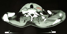
![Hodgkin lymphoma-Gross mesenteric mass lesions consistent with lymphadenopathy. Spleen is also enlarged.[2]](/images/3/39/Hodgkin%27s_lymphoma_CT_01.jpg)
Hodgkin lymphoma-Gross mesenteric mass lesions consistent with lymphadenopathy. Spleen is also enlarged.[2]
-
![Hodgkin lymphoma-Gross mesenteric mass lesions consistent with lymphadenopathy. Spleen is also enlarged.[2]](/images/4/4e/Hodgkin%27s_lymphoma_CT_02.jpg)
Hodgkin lymphoma-Gross mesenteric mass lesions consistent with lymphadenopathy. Spleen is also enlarged.[2]
-
![Hodgkin lymphoma-Gross mesenteric mass lesions consistent with lymphadenopathy. Spleen is also enlarged.[2]](/images/7/75/Hodgkin%27s_lymphoma_CT_03.jpg)
Hodgkin lymphoma-Gross mesenteric mass lesions consistent with lymphadenopathy. Spleen is also enlarged.[2]
.jpg)
![Hodgkin lymphoma-Gross mesenteric mass lesions consistent with lymphadenopathy. Spleen is also enlarged.[2]](/index.php/File:Hodgkin%27s_lymphoma_CT_01.jpg)
![Hodgkin lymphoma-Gross mesenteric mass lesions consistent with lymphadenopathy. Spleen is also enlarged.[2]](/index.php/File:Hodgkin%27s_lymphoma_CT_02.jpg)
![Hodgkin lymphoma-Gross mesenteric mass lesions consistent with lymphadenopathy. Spleen is also enlarged.[2]](/index.php/File:Hodgkin%27s_lymphoma_CT_03.jpg)
References
- ↑ Hodgkin-lymphoma. Canadian Cancer Society. http://www.cancer.ca/en/cancer-information/cancer-type/hodgkin-lymphoma/diagnosis/?region=ab Accessed on September 11, 2015
- ↑ 2.0 2.1 2.2 Image courtesy of Dr Chris O'DonnellRadiopaedia(original file ‘’here’’). Creative Commons BY-SA-NC