Hernia
| Hernia | |
 | |
|---|---|
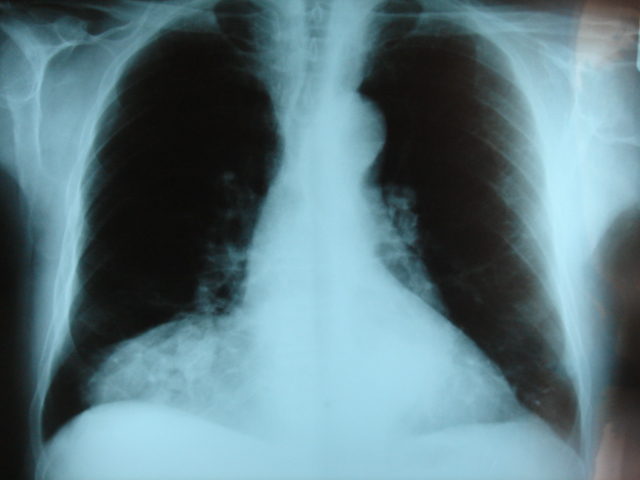
| Frontal chest X-ray showing a hernia of Morgagni | |
| ICD-10 | K40-K46 |
| ICD-9 | 550-553 |
| MedlinePlus | 000960 |
|
Hernia Microchapters |
|
Diagnosis |
|---|
|
Treatment |
|
Case Studies |
|
Hernia On the Web |
|
American Roentgen Ray Society Images of Hernia |
For the WikiPatient page for this topic, click here
Editor-In-Chief: C. Michael Gibson, M.S., M.D. [1] Associate Editor-In-Chief: Mohammed A. Sbeih, M.D. [2]
Overview
A hernia is a protrusion of a tissue, structure, or part of an organ through the muscular tissue or the membrane by which it is normally contained. The hernia has 3 parts: the orifice through which it herniates, the hernial sac, and its contents. The contents, usually portions of intestine or abdominal fatty tissue, are enclosed in the thin membrane that naturally lines the inside of the cavity. a Hernia has a potential risk of having its blood supply cut off (becoming strangulated), and the contents may become necrotic due to the lack of O2 supply.
A hernia may be likened to a failure in the sidewall of a pneumatic tire. The tire's inner tube behaves like the organ and the side wall like the body cavity wall providing the restraint. A weakness in the sidewall allows a bulge to develop, which can become a split, allowing the inner tube to protrude, and leading to the eventual failure of the tire.
Treatment
It is generally advisable to repair hernias in a timely fashion, in order to prevent complications such as organ dysfunction, gangrene, and multiple organ dysfunction syndrome. Most abdominal hernias can be surgically repaired, and recovery rarely requires long-term changes in lifestyle. Uncomplicated hernias are principally repaired by pushing back, or "reducing", the herniated tissue, and then mending the weakness in muscle tissue (an operation called herniorrhaphy). If complications have occurred, the surgeon will check the viability of the herniated organ, and resect it if necessary. Modern muscle reinforcement techniques involve synthetic materials (a mesh prosthesis) that avoid over-stretching of already weakened tissue (as in older, but still useful methods). The mesh is placed over the defect, and sometimes staples are used to keep the mesh in place. Increasingly, some repairs are performed through laparoscopes.
Many patients are managed through surgical daycare centers, and are able to return to work within a week or two, while heavy activities are prohibited for a longer period. Surgical complications have been estimated to be up to 10%, but most of them can be easily addressed. They include surgical site infections, nerve and blood vessel injuries, injury to nearby organs, and hernia recurrence.
Generally, the use of external devices to maintain reduction of the hernia without repairing the underlying defect (such as hernia trusses, trunks, belts, etc.), is not advised. Exceptions are uncomplicated incisional hernias that arise shortly after the operation (should only be operated after a few months), or inoperable patients.
It is essential that the hernia not be further irritated by carrying out strenuous labour.
Individual hernias
A sportman's hernia is a syndrome characterized by chronic groin pain in athletes and a dilated superficial ring of the inguinal canal, although a true hernia is not present.
Inguinal hernia
- Main article: inguinal hernia.

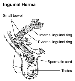
By far the most common hernias (up to 75% of all abdominal hernias) are the so-called inguinal hernias. For a thorough understanding of inguinal hernias, much insight is needed in the anatomy of the inguinal canal. Inguinal hernias are further divided into the more common indirect inguinal hernia (2/3, depicted here), in which the inguinal canal is entered via a congenital weakness at its entrance (the internal inguinal ring), and the direct inguinal hernia type (1/3), where the hernia contents push through a weak spot in the back wall of the inguinal canal. Inguinal hernias are more common in men than women while femoral hernias are more common in women.
Femoral hernia
- Main article: femoral hernia.
Femoral hernias occur just below the inguinal ligament, when abdominal contents pass into the weak area at the posterior wall of the femoral canal. They can be hard to distinguish from the inguinal type (especially when ascending cephalad): however, they generally appear more rounded, and, in contrast to inguinal hernias, there is a strong female preponderance in femoral hernias. The incidence of strangulation in femoral hernias is high. Repair techniques are similar for femoral and inguinal hernia.
Umbilical hernia
- Main article: umbilical hernia.
Umbilical hernias are especially common in infants of African descent, and occur more in boys. They involve protrusion of intraabdominal contents through a weakness at the site of passage of the umbilical cord through the abdominal wall. These hernias often resolve spontaneously. Umbilical hernias in adults are largely acquired, and are more frequent in obese or pregnant women. Abnormal decussation of fibers at the linea alba may contribute.
Incisional hernia
- Main article: incisional hernia.
An incisional hernia occurs when the defect is the result of an incompletely healed surgical wound. When these occur in median laparotomy incisions in the linea alba, they are termed ventral hernias. These can be the most frustrating and difficult to treat, as the repair utilizes already attenuated tissue.
Diaphragmatic hernia
- Main article: diaphragmatic hernia

Higher in the abdomen, an (internal) "diaphragmatic hernia" results when part of the stomach or intestine protrudes into the chest cavity through a defect in the diaphragm.
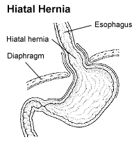
A hiatus hernia is a particular variant of this type, in which the normal passageway through which the esophagus meets the stomach (esophageal hiatus) serves as a functional "defect", allowing part of the stomach to (periodically) "herniate" into the chest. Hiatus hernias may be either "sliding," in which the gastroesophageal junction itself slides through the defect into the chest, or non-sliding (also known as para-esophageal), in which case the junction remains fixed while another portion of the stomach moves up through the defect. Non-sliding or para-esophageal hernias can be dangerous as they may allow the stomach to rotate and obstruct. Repair is usually advised.

A congenital diaphragmatic hernia is a distinct problem, occurring in up to 1 in 2000 births, and requiring pediatric surgery. Intestinal organs may herniate through several parts of the diaphragm, posterolateral (in Bochdalek's triangle, resulting in Bochdalek's hernia), or anteromedial-retrosternal (in the cleft of Larrey/Morgagni's foramen, resulting in Morgagni-Larrey hernia, or Morgagni's hernia).
Other types of hernia
Since many organs or parts of organs can herniate through many orifices, it is very difficult to give an exhaustive list of hernias, with all synonyms and eponyms. The above article deals mostly with "visceral hernias", where the herniating tissue arises within the abdominal cavity. Other hernia types and unusual types of visceral hernias are listed below, in alphabetical order:
- Brain hernia: herniation of part of the brain because of excessive intracranial pressure. This may be a life-threatening condition, especially if the brain stem (responsible for some important vital signs) is involved.
- Cooper's hernia: A femoral hernia with two sacs, the first being in the femoral canal, and the second passing through a defect in the superficial fascia and appearing immediately beneath the skin.
- epigastric hernia: hernia through the linea alba above the umbilicus.
- Littre's hernia: hernia involving a Meckel's diverticulum. It is named after French anatomist Alexis Littre (1658-1726).
- Lumbar hernia: hernia in the lumbar region (not to be confused with a lumbar disc hernia), contains following entities:
1. Petit's hernia - hernia through Petit's triangle (inferior lumbar triangle). It is named after French surgeon Jean Louis Petit (1674-1750).
2. Grynfeltt's hernia - hernia through Grynfeltt-Lesshaft triangle (superior lumbar triangle). It is named after physician Joseph Grynfeltt (1840-1913).
- obturator hernia: hernia through obturator canal.
- pantaloon hernia: a combined direct and indirect hernia, when the hernial sac protrudes on either side of the inferior epigastric vessels.
- perineal hernia: A perineal hernia protrudes through the muscles and fascia of the perineal floor. It may be primary but usually, is acquired following perineal prostatectomy, abdominoperineal resection of the rectum, or pelvic exenteration.
- properitoneal hernia: rare hernia located directly above the peritoneum, for example, when part of an inguinal hernia projects from the deep inguinal ring to the preperitoneal space.
- Richter's hernia: strangulated hernia involving only one sidewall of the bowel, which can result in bowel perforation through ischaemia without causing bowel obstruction or any of its warning signs. It is named after German surgeon August Gottlieb Richter (1742-1812).
- sliding hernia: occurs when an organ drags along part of the peritoneum, or, in other words, the organ is part of the hernia sac. The colon and the urinary bladder are often involved. The term also frequently refers to sliding hernias of the stomach.
- sciatic hernia: this hernia in the greater sciatic foramen most commonly presents as an uncomfortable mass in the gluteal area. Bowel obstruction may also occur. This type of hernia is only a rare cause of sciatic neuralgia.
- Spigelian hernia, also known as spontaneous lateral ventral hernia.
- Velpeau hernia: a hernia in the groin in front of the femoral blood vessels.
- spinal disc herniation, or "herniated nucleus pulposus": a condition where the central weak part of the intervertebral disc (nucleus pulposus, which helps absorb shocks to our spine), herniates through the fibrous band (annulus fibrosus) by which it is normally bound. This usually occurs low in the back at the lumbar or lumbo-sacral level and can cause back pain which usually radiates well into the thigh or leg. When the sciatic nerve is involved, the symptom complex is called sciatica. Herniation can occur in the cervical vertebrae too. A nucleoplasty is an operation to repair the herniation.
- Traumatic abdominal wall hernia: herniation of viscera occurring after a force is applied to the abdomen in a patient without preexisting abdominal hernia, resulting in disruption of muscles and fascia while maintaining skin continuity.
Complications
Complications may arise post-operation, including rejection of the mesh that is used to repair the hernia. In the event of a mesh rejection, the mesh will very likely need to be removed. Mesh rejection can be detected by obvious, sometimes localised swelling and pain around the mesh area. Continuous discharge from the scar is likely for a while after the mesh has been removed.
An untreated hernia may complicate by:
- Inflammation
- Irreducibilty
- Obstruction
- Strangulation
- Hydrocele of the hernial sac
References
- Surgical recall, 2nd edition, by Lorne. H. Blackbourne, published by Lippincott Williams & Wilkins
- Sabiston textbook of surgery, 17th edition, Townsend et.al.(e.d.), Elsevier-Saunders
External links
ca:Hèrnia de:Hernie ko:탈장 it:Ernia he:בקע nl:Hernia no:Brokk nn:brokk sr:Брух fi:Tyrä sv:Bråck