Hernia classification
|
Hernia Microchapters |
|
Diagnosis |
|---|
|
Treatment |
|
Case Studies |
|
Hernia classification On the Web |
|
American Roentgen Ray Society Images of Hernia classification |
Editor-In-Chief: C. Michael Gibson, M.S., M.D. [1]
Classification
Classification Based on Anatomic Location
Hernias can be classified according to their anatomical location:
Examples include:
- Abdominal hernias.
- Diaphragmatic hernias and hiatal hernias (for example, paraesophageal hernia of the stomach).
- Pelvic hernias, for example, obturator hernia.
- Hernias of the nucleus pulposus of the intervertebral discs.
- Intracranial hernias.
Sportsman's Hernia
A sportman's hernia is a syndrome characterized by chronic groin pain in athletes and a dilated superficial ring of the inguinal canal, although a true hernia is not present.
Inguinal Hernia
- Main article: inguinal hernia.

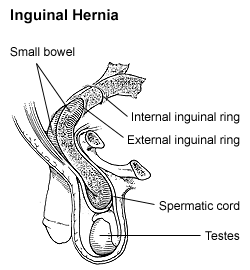
By far the most common hernias (up to 75% of all abdominal hernias) are the so-called inguinal hernias. For a thorough understanding of inguinal hernias, much insight is needed in the anatomy of the inguinal canal. Inguinal hernias are further divided into the more common indirect inguinal hernia (2/3, depicted here), in which the inguinal canal is entered via a congenital weakness at its entrance (the internal inguinal ring), and the direct inguinal hernia type (1/3), where the hernia contents push through a weak spot in the back wall of the inguinal canal. Inguinal hernias are more common in men than women while femoral hernias are more common in women.
Femoral Hernia
- Main article: femoral hernia.
Femoral hernias occur just below the inguinal ligament, when abdominal contents pass into the weak area at the posterior wall of the femoral canal. They can be hard to distinguish from the inguinal type (especially when ascending cephalad): however, they generally appear more rounded, and, in contrast to inguinal hernias, there is a strong female preponderance in femoral hernias. The incidence of strangulation in femoral hernias is high. Repair techniques are similar for femoral and inguinal hernia.
Umbilical Hernia
- Main article: umbilical hernia.
Umbilical hernias are especially common in infants of African descent, and occur more in boys. They involve protrusion of intraabdominal contents through a weakness at the site of passage of the umbilical cord through the abdominal wall. These hernias often resolve spontaneously. Umbilical hernias in adults are largely acquired, and are more frequent in obese or pregnant women. Abnormal decussation of fibers at the linea alba may contribute.
Incisional Hernia
- Main article: incisional hernia.
An incisional hernia occurs when the defect is the result of an incompletely healed surgical wound. When these occur in median laparotomy incisions in the linea alba, they are termed ventral hernias. These can be the most frustrating and difficult to treat, as the repair utilizes already attenuated tissue.
Diaphragmatic Hernia
- Main article: diaphragmatic hernia

Higher in the abdomen, an (internal) "diaphragmatic hernia" results when part of the stomach or intestine protrudes into the chest cavity through a defect in the diaphragm.
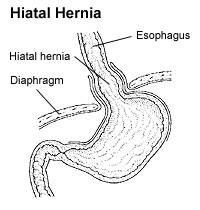
A hiatus hernia is a particular variant of this type, in which the normal passageway through which the esophagus meets the stomach (esophageal hiatus) serves as a functional defect, allowing part of the stomach to (periodically) herniate into the chest. Hiatus hernias may be either sliding, in which the gastroesophageal junction itself slides through the defect into the chest, or non-sliding (also known as para-esophageal), in which case the junction remains fixed while another portion of the stomach moves up through the defect. Non-sliding or para-esophageal hernias can be dangerous as they may allow the stomach to rotate and obstruct. Repair is usually advised.

A congenital diaphragmatic hernia is a distinct problem, occurring in up to 1 in 2000 births, and requiring pediatric surgery. Intestinal organs may herniate through several parts of the diaphragm, posterolateral (in Bochdalek's triangle, resulting in Bochdalek's hernia), or anteromedial-retrosternal (in the cleft of Larrey/Morgagni's foramen, resulting in Morgagni-Larrey hernia, or Morgagni's hernia).
Other Types of Hernia
Since many organs or parts of organs can herniate through many orifices, it is very difficult to give an exhaustive list of hernias, with all synonyms and eponyms. The above article deals mostly with "visceral hernias", where the herniating tissue arises within the abdominal cavity. Other hernia types and unusual types of visceral hernias are listed below, in alphabetical order:
- Brain hernia: herniation of part of the brain because of excessive intracranial pressure. This may be a life-threatening condition, especially if the brain stem (responsible for some important vital signs) is involved.
- Cooper's hernia: a femoral hernia with two sacs, the first being in the femoral canal, and the second passing through a defect in the superficial fascia and appearing immediately beneath the skin.
- Epigastric hernia: hernia through the linea alba above the umbilicus.
- Littre's hernia: hernia involving a Meckel's diverticulum. It is named after French anatomist Alexis Littre (1658-1726).
- Lumbar hernia: hernia in the lumbar region (not to be confused with a lumbar disc hernia), contains following entities:
- Petit's hernia - Hernia through Petit's triangle (inferior lumbar triangle). It is named after French surgeon Jean Louis Petit (1674-1750).
- Grynfeltt's hernia - Hernia through Grynfeltt-Lesshaft triangle (superior lumbar triangle). It is named after physician Joseph Grynfeltt (1840-1913).
- Obturator hernia: hernia through obturator canal.
- Pantaloon hernia: a combined direct and indirect hernia, when the hernial sac protrudes on either side of the inferior epigastric vessels.
- Perineal hernia: a perineal hernia protrudes through the muscles and fascia of the perineal floor. It may be primary but usually, is acquired following perineal prostatectomy, abdominoperineal resection of the rectum, or pelvic exenteration.
- Properitoneal hernia: rare hernia located directly above the peritoneum, for example, when part of an inguinal hernia projects from the deep inguinal ring to the preperitoneal space.
- Richter's hernia: strangulated hernia involving only one sidewall of the bowel, which can result in bowel perforation through ischaemia without causing bowel obstruction or any of its warning signs. It is named after German surgeon August Gottlieb Richter (1742-1812).
- Sliding hernia: occurs when an organ drags along part of the peritoneum, or, in other words, the organ is part of the hernia sac. The colon and the urinary bladder are often involved. The term also frequently refers to sliding hernias of the stomach.
- Siatic hernia: this hernia in the greater sciatic foramen most commonly presents as an uncomfortable mass in the gluteal area. Bowel obstruction may also occur. This type of hernia is only a rare cause of sciatic neuralgia.
- Spigelian hernia, also known as spontaneous lateral ventral hernia.
- Velpeau hernia: a hernia in the groin in front of the femoral blood vessels.
- Spinal disc herniation, or herniated nucleus pulposus: a condition where the central weak part of the intervertebral disc (nucleus pulposus, which helps absorb shocks to our spine), herniates through the fibrous band (annulus fibrosus) by which it is normally bound. This usually occurs low in the back at the lumbar or lumbo-sacral level and can cause back pain which usually radiates well into the thigh or leg. When the sciatic nerve is involved, the symptom complex is called sciatica. Herniation can occur in the cervical vertebrae too. A nucleoplasty is an operation to repair the herniation.
- Traumatic abdominal wall hernia: herniation of viscera occurring after a force is applied to the abdomen in a patient without preexisting abdominal hernia, resulting in disruption of muscles and fascia while maintaining skin continuity.
Classification Based on Characteristics
Each of the above hernias may be characterized by several aspects:
- Congenital or acquired: congenital hernias occur prenatally or in the first year(s) of life, and are caused by a congenital defect, whereas acquired hernias develop later on in life. However, this may be on the basis of a locus minoris resistentiae (Lat. place of least resistance) that is congenital, but only becomes symptomatic later on in life, when degeneration and increased stress (for example, increased abdominal pressure from coughing in COPD) provoke the hernia.
- Complete or incomplete: for example, the stomach may partially herniate into the chest, or completely.
- Internal or external: external ones herniate to the outside world, whereas internal hernias protrude from their normal compartment to another (for example, mesenteric hernias).
- Intraparietal hernia: hernia that does not reach all the way to the subcutis, but only to the musculoaponeurotic layer. An example is a Spigelian hernia. Intraparietal hernias may produces less obvious bulging, and may be less easily detected on clinical examination.
- Bilateral: in this case, simultaneous repair may be considered, sometimes even with a giant prosthetic reinforcement.
- Irreducible (also known as incarcerated): the hernial contents cannot be returned to their normal site with simple manipulation.
If irreducible, hernias can develop several complications (hence, they can be complicated or uncomplicated):
- Strangulation: pressure on the hernial contents may compromise blood supply (especially veins, with their low pressure, are sensitive, and venous congestion often results) and cause ischemia, and later necrosis and gangrene, which may become fatal.
- Obstruction: for example, when a part of the bowel herniates, bowel contents can no longer pass the obstruction. This results in cramps, and later on vomiting, ileus, absence of flatus and absence of defecation. These signs mandate urgent surgery.
- Another complication arises when the herniated organ itself, or surrounding organs start dysfunctioning (for example, sliding hernia of the stomach causing heartburn, lumbar disc hernia causing sciatic nerve pain, etc.)