Hepatocellular carcinoma pathophysiology
| https://https://www.youtube.com/watch?v=zv04qtEM8qw%7C350}} |
|
Hepatocellular carcinoma Microchapters |
|
Differentiating Hepatocellular carcinoma from other Diseases |
|---|
|
Diagnosis |
|
Treatment |
|
Case Studies |
|
Hepatocellular carcinoma pathophysiology On the Web |
|
American Roentgen Ray Society Images of Hepatocellular carcinoma pathophysiology |
|
Risk calculators and risk factors for Hepatocellular carcinoma pathophysiology |
Editor-In-Chief: C. Michael Gibson, M.S., M.D. [1]Associate Editor(s)-in-Chief: Dildar Hussain, MBBS [2] Parminder Dhingra, M.D. [3] Mohamad Alkateb, MBBCh [4]
Overview
The exact pathogenesis of hepatocellular carcinoma HCC is not fully understood. It is thought that HCC is mediated by either HBV infection, HCV infection, underlying cirrhotic liver disease, inflammation, necrosis and fibrosis. On microscopic histopathological analysis, large polygonal tumours cells with graunular eosinophilic cytoplasm or layered dense collagen bundles are characteristic findings of hepatocellular carcinoma.
Pathophysiology
Pathogenesis
- The exact pathogenesis of hepatocellular carcinoma is not fully understood. HCC arises from precancerous lesions and well-differentiated HCC further progresses to a less differentiated form. There is still an enormous demand for the explanation of objective morphological, phenotypic and genetic markers for the progression of HCC.
- It is thought that hepatocellular carcinoma is caused by:[1][2]
- Chronic exposure of liver insult due to HBV infection,HCV infection,aflatoxin,nonalcoholic steatohepatitis (NASH) causes damage to the hepatocytes induces a viscious circle of destruction and regeneration of hepatocytes which finally causes the activation of stellate cells and hepatocyte senescence which contributes in the development of cirrhosis.
- As a result of the genomic unstability the initiation of HCC occurs, step wise multiplication of different genetic events that lead to tumor progression and metastases are:
- Gene rearrangements
- Somatic mutations
- Copy number alterations
- Epigenetic changes
- Growth factor pathway alterations
- Molecular pathway alterations
To review the pathogenesis of HBV infection, click here.
To review the pathogenesis of HCV infection click here.
To review the pathogenesis of hepatic cirrhosis, click here.
Genetics
- As of now there is no significant manifestation of an ordered cycle of genomic events leading to hepatocarcinogenesis. The pattern of genomic transformations exhibit huge variations often between two different HCCs from a single patient.
- Hepatocellular carcinoma is most commonly implicated with underlying chronic hepatitis and liver cirrhosis, however differen genes have been associated with the pathogenesis of the HCC, which are further divided into four major groups:[2][3]
- Genes regulating DNA damage response
- Genes involved in cell cycle control
- Genes involved in growth inhibition and apoptosis
- Genes responsible for cell–cell interaction and signal transduction.
- Genes involved in the pathogenesis of hepatocellular carcinoma include:
Associated Conditions
The associated conditions with hepatocellular carcinoma are
- HBV infection
- HCV infection
- Underlying cirrhotic liver disease
- Inflammation
- Necrosis and fibrosis of the liver
Microscopic Pathology
- On microscopic histopathological analysis, large polygonal tumours cells with graunular eosinophilic cytoplasm or layered dense collagen bundles are characteristic findings of hepatocellular carcinoma.
Gross Pathology
On gross pathology, hepatocellular carcinoma has the following characteristic findings:[4]
- Nodular or diffusely infiltrative.
- The nodular type may be unifocal (large mass) or multifocal (when developed as a complication of cirrhosis). Tumor nodules are round to oval, grey or green (if the tumor produces bile), well circumscribed but not encapsulated.
- In about fifty percent of the cases, the tumors are multifocal where as some authors have suggested it to be around 75 percent.
- The portal vein is infiltrated by the poorly circumcised diffused type and the hepatic veins are rarely infiltrated.
- Pale in relation to surrounding liver or green (due to bile secretion)
-
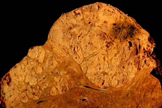
Hepatocelluler carcinoma. The image shows a longitudinal slice taken through the full length of the liver.
(Courtesy of Ed Uthman, MD) -

The tumor is at the top, cirrhotic liver at the bottom, and a fibrous reaction in between. Hepatocellular carcinomas can have a variety of gross patterns, including multinodular / multifocal, such as this one.
(Courtesy of Ed Uthman, MD)


Microscopic Pathology
On microscopic histopathological analysis, hepatocellular carcinoma has the following characteristic findings:
- Large polygonal tumours cells with:
- Graunular eosinophilic cytoplasm
- Low NC ratio
- Layered dense collagen bundles [5]
Videos
{{#ev:youtube|qsR92joabBg}}
References
- ↑ Röcken C, Carl-McGrath S (2001). "Pathology and pathogenesis of hepatocellular carcinoma". Dig Dis. 19 (4): 269–78. doi:10.1159/000050693. PMID 11935086.
- ↑ 2.0 2.1 Dhanasekaran R, Bandoh S, Roberts LR (2016). "Molecular pathogenesis of hepatocellular carcinoma and impact of therapeutic advances". F1000Res. 5. doi:10.12688/f1000research.6946.1. PMC 4870992. PMID 27239288.
- ↑ Li M, Zhao H, Zhang X, Wood LD, Anders RA, Choti MA, Pawlik TM, Daniel HD, Kannangai R, Offerhaus GJ, Velculescu VE, Wang L, Zhou S, Vogelstein B, Hruban RH, Papadopoulos N, Cai J, Torbenson MS, Kinzler KW (2011). "Inactivating mutations of the chromatin remodeling gene ARID2 in hepatocellular carcinoma". Nat. Genet. 43 (9): 828–9. doi:10.1038/ng.903. PMC 3163746. PMID 21822264.
- ↑ Reynolds AR, Furlan A, Fetzer DT, Sasatomi E, Borhani AA, Heller MT, Tublin ME (2015). "Infiltrative hepatocellular carcinoma: what radiologists need to know". Radiographics. 35 (2): 371–86. doi:10.1148/rg.352140114. PMID 25763723.
- ↑ "Hepatocellular carcinoma".