Cyanosis pathophysiology
|
Cyanosis Microchapters |
|
Diagnosis |
|---|
|
Treatment |
|
Case Studies |
|
Cyanosis pathophysiology On the Web |
|
American Roentgen Ray Society Images of Cyanosis pathophysiology |
|
Risk calculators and risk factors for Cyanosis pathophysiology |
Editor-In-Chief: C. Michael Gibson, M.S., M.D. [1]; Associate Editor(s)-in-Chief: Chandrakala Yannam, MD [2]
Overview
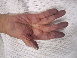
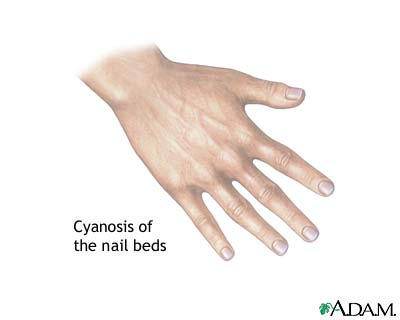
Cyanosis is a bluish or purplish discoloration of the skin and mucous membranes. Two mechanisms involved in the development of cyanosis, Systemic arterial oxygen desaturation and increased oxygen absorption by tissues. Cyanosis is evident when arterial oxygen desaturation falls below 85% or the concentration of deoxygenated hemoglobin (Hb) is below 5 gm/dl. Several factors can affect the appearance of cyanosis includes skin pigmentation, Hemoglobin (Hb) levels, oxygen affinity to the hemoglobin (Hb).
Pathophysiology
- Cyanosis is a bluish or purplish discoloration of the skin and mucous membranes.
- Appearance of cyanosis depends on the absolute amount of deoxygenated hemoglobin(Hb) present in the blood rather than the ratio of reduced hemoglobin (Hb) to oxygenated hemoglobin (Hb).[1][2]
- According to Lundsgaard and Van Slyke (1923), as well as subsequent investigators, cyanosis is evident when the subpapillary capillaries contain from 4 to 6 gm/dl of deoxygenated hemoglobin and oxygenation of hemoglobin or oxygen saturation falls below 85%.[3]
- Cyanosis occurs due to following mechanisms:
- Central cyanosis:
- Central cyanosis is caused by reduced arterial oxygen saturation or the presence of abnormal hemoglobin derivatives (methemoglobin or sulfhemoglobin).
- Central cyanosis is evident when the systemic arterial deoxygenated hemoglobin concentration in the blood exceeds 5 g/dL (oxygen saturation ≤85 percent).
- The increased amount of deoxygenated hemoglobin is the result of either increased amount of venous admixture or reduced capillary arterial oxygen tension.
- Peripheral cyanosis:
- In peripheral cyanosis, systemic arterial oxygen saturation is normal.
- Increased oxygen extraction by tissues causes wide systemic arteriovenous oxygen difference and increased deoxygenated blood on the venous side of the capillary beds.
- The increased oxygen extraction by tissues results from the sluggish movement of blood through the capillary circulation.
- Causes for reduced blood flow through capillary circulation include:
- Vasoconstriction caused by exposure to cold
- Venous obstruction
- Elevated venous pressure
- Hyperviscosity (Polycythemia)
- Low cardiac output
- Vasomotor instability

- Factors can affect the development of cyanosis:
- Hemoglobin concentration
- Skin pigmentation
- Presence of abnormal hemoglobins interfering with oxygen affinity
- Lighting conditions
- Factors can affect the development of cyanosis:
Genetics, Associated Conditions, Gross Pathology, Microscopic Pathology

By James Heilman, MD (Own work) [CC BY-SA 3.0 (https://creativecommons.org/licenses/by-sa/3.0)], via Wikimedia Commons
For the detailed information of the genetics, associated conditions, gross and microscopic pathological features associated with conditions causing cyanosis, click the links below.
- Tetralogy of Fallot
- Tricuspid atresia
- Ebstein's anomaly
- Tricuspid stenosis
- Transposition of great arteries (TGA)
- Pulmonary stenosis
- Truncus arteriosus
- TAPVC
- Coarctation of aorta
- Aortic stenosis
- Eisenmenger's syndrome
- HLHS (Spectrum of hypoplastic left heart syndrome)
- Left-sided heart failure
- Acute chest syndrome
- Pneumothorax
- Foreign body aspiration
- Carbon monoxide poisoning
- Hydrogen cyanide poisoning
- Croup
- Bacterial tracheitis
- Hemothorax
- Pneumonia
- Asthma
- COPD
- Bronchiolitis
- Respiratory distress syndrome (Hyaline membrane disease)
- Empyema
- Pleural effusion
- Cystic fibrosis
- Atelectasis
- Bronchopulmonary dysplasia
- Alveolar capillary dysplasia
- Pulmonary embolism
- Pulmonary hypertension
- High Altitude
- Intracranial hemorrhage
- Seizures
- Choanal atresia
- Micrognathia or retrognathia
- Laryngomalacia
- Congenital diaphragmatic hernia
- Myasthenia gravis
- Apnea of prematurity
- Obstructive sleep apnea
- Pulmonary edema
- Pulmonary hemorrhage
- Pulmonary arteriovenous malformation
- Methemoglobinemia (congenital or acquired)
- Sulfhemoglobinemia
- Polycythemia vera
- Disseminated intravascular coagulation
- Shock
- Sepsis
- Amniotic fluid embolism
- Cold exposure
- Acrocyanosis
- Raynaud's phenomenon
- Raynaud's disease
- Peripheral vascular disease
- Buergers disease
- Deep vein thrombosis
- Superior vena cava syndrome
References
- ↑ Blount SG (May 1971). "Cyanosis: pathophysiology and differential diagnosis". Prog Cardiovasc Dis. 13 (6): 595–605. PMID 4933007.
- ↑ GERACI JE, WOOD EH (July 1951). "The relationship of the arterial oxygen saturation to cyanosis". Med. Clin. North Am. 1: 1185–1202. PMID 13098533.
- ↑ Lundsgaard C (September 1919). "STUDIES ON CYANOSIS : I. PRIMARY CAUSES OF CYANOSIS". J. Exp. Med. 30 (3): 259–69. PMC 2126682. PMID 19868357.
- ↑ Adeyinka A, Kondamudi NP. PMID 29489181. Missing or empty
|title=(help)