Chickenpox physical examination
Editor-In-Chief: C. Michael Gibson, M.S., M.D. [1]
Michael Maddaleni, B.S.
|
Chickenpox Microchapters |
|
Diagnosis |
|---|
|
Treatment |
|
Case Studies |
|
Chickenpox physical examination On the Web |
|
American Roentgen Ray Society Images of Chickenpox physical examination |
|
Risk calculators and risk factors for Chickenpox physical examination |
Overview
The diagnosis of varicella is primarily clinical. In a non-immunized individual with the appropriate appearing rash occurring in "crops", no further investigation would normally be undertaken. For further investigation, examination of the fluid within the vesicles, or by testing blood for evidence of an acute immunologic response. Vesicle fluid can be examined with a Tsanck smear, or better with examination for direct fluorescent antibody. The fluid can also be "cultured", whereby attempts are made to grow the virus from a fluid sample. Blood tests can be used to identify a response to acute infection (IgM) or previous infection and subsequent immunity (IgE).[1]. Prenatal diagnosis of foetal varicella infection can be performed using ultrasound, though a delay of 5 weeks following primary maternal infection is advised. A PCR (DNA) test of the mother's amniotic fluid can also be performed, though the risk of spontaneous abortion due to the amniocentesis procedure is higher than the risk of the baby developing foetal varicella syndrome.
Physical Examination
General appearnace
Vitals
Skin
HEENT
Neck
Lungs
Heart
Abdomen
Extremities
Neuromuscular
Gallery
-
![Chickenpox lesions on the skin of this patient's left breast and arm on day 6 of the illness. From Public Health Image Library (PHIL). [2]](/images/9/95/Chickenpox36.jpeg)
Chickenpox lesions on the skin of this patient's left breast and arm on day 6 of the illness. From Public Health Image Library (PHIL). [2]
-
![Chickenpox lesions on the skin of this patient's back and buttocks at day 6 of the illness. From Public Health Image Library (PHIL). [2]](/images/8/8e/Chickenpox35.jpeg)
Chickenpox lesions on the skin of this patient's back and buttocks at day 6 of the illness. From Public Health Image Library (PHIL). [2]
-
![Chickenpox lesions on the skin of this patient's breasts, arms, and torso at day 6 of the illness. From Public Health Image Library (PHIL). [2]](/images/7/71/Chickenpox34.jpeg)
Chickenpox lesions on the skin of this patient's breasts, arms, and torso at day 6 of the illness. From Public Health Image Library (PHIL). [2]
-
![Patient with cervical skin lesions caused by chickenpox. From Public Health Image Library (PHIL). [2]](/images/c/c7/Chickenpox33.jpeg)
Patient with cervical skin lesions caused by chickenpox. From Public Health Image Library (PHIL). [2]
-
![4-month old infant with skin lesions on his brow ridge due to chickenpox. From Public Health Image Library (PHIL). [2]](/images/0/03/Chickenpox32.jpeg)
4-month old infant with skin lesions on his brow ridge due to chickenpox. From Public Health Image Library (PHIL). [2]
-
![Patient had presented with chickenpox demonstrating the typical rash on day eight. From Public Health Image Library (PHIL). [2]](/images/8/84/Chickenpox29.jpeg)
Patient had presented with chickenpox demonstrating the typical rash on day eight. From Public Health Image Library (PHIL). [2]
-
![Patient developed palatal mucosal lesions due to chickenpox. From Public Health Image Library (PHIL). [2]](/images/1/18/Chickenpox28.jpeg)
Patient developed palatal mucosal lesions due to chickenpox. From Public Health Image Library (PHIL). [2]
-
![Vaccine recipient developed a secondary herpes infection adjacent to the vaccination site. From Public Health Image Library (PHIL). [2]](/images/3/3e/Chickenpox27.jpeg)
Vaccine recipient developed a secondary herpes infection adjacent to the vaccination site. From Public Health Image Library (PHIL). [2]
-
![Pustulovesicular rash represents a generalized herpes outbreak due to the Varicella-zoster virus (VZV) pathogen. From Public Health Image Library (PHIL). [2]](/images/6/6e/Chickenpox26.jpeg)
Pustulovesicular rash represents a generalized herpes outbreak due to the Varicella-zoster virus (VZV) pathogen. From Public Health Image Library (PHIL). [2]
-
![Case of chickenpox. From Public Health Image Library (PHIL). [2]](/images/e/e2/Chickenpox24.jpeg)
Case of chickenpox. From Public Health Image Library (PHIL). [2]
-
![Case of chickenpox. From Public Health Image Library (PHIL). [2]](/images/6/6c/Chickenpox23.jpeg)
Case of chickenpox. From Public Health Image Library (PHIL). [2]
-
![Case of chickenpox. From Public Health Image Library (PHIL). [2]](/images/5/5a/Chickenpox22.jpeg)
Case of chickenpox. From Public Health Image Library (PHIL). [2]
-
![Chickenpox lesions on a patient’s back, which were displaying the characteristic “cropping” distribution, or manifesting themselves in clusters. From Public Health Image Library (PHIL). [2]](/images/4/47/Chickenpox21.jpeg)
Chickenpox lesions on a patient’s back, which were displaying the characteristic “cropping” distribution, or manifesting themselves in clusters. From Public Health Image Library (PHIL). [2]
-
![Posterior view of a hospitalized man's neck, back and shoulders, who’d been assigned a bed in a smallpox ward, due to an initially misdiagnosed illness, which turned out to be chickenpox. From Public Health Image Library (PHIL). [2]](/images/e/e9/Chickenpox20.jpeg)
Posterior view of a hospitalized man's neck, back and shoulders, who’d been assigned a bed in a smallpox ward, due to an initially misdiagnosed illness, which turned out to be chickenpox. From Public Health Image Library (PHIL). [2]
-
![View of a patient’s thighs and upper legs, who’d been diagnosed with chickenpox. From Public Health Image Library (PHIL). [2]](/images/2/21/Chickenpox19.jpeg)
View of a patient’s thighs and upper legs, who’d been diagnosed with chickenpox. From Public Health Image Library (PHIL). [2]
-
![Pathologic changes seen on the surface of the right unilateral side of this elderly male patient’s tongue and chin, represent a herpes outbreak due to the Varicella-zoster virus (VZV) pathogen. From Public Health Image Library (PHIL). [2]](/images/b/b8/Chickenpox18.jpeg)
Pathologic changes seen on the surface of the right unilateral side of this elderly male patient’s tongue and chin, represent a herpes outbreak due to the Varicella-zoster virus (VZV) pathogen. From Public Health Image Library (PHIL). [2]
-
![Viewed from above, this image depicts a smallpox scab (left), and chickenpox scab (right) as a demonstration in comparative morphology. From Public Health Image Library (PHIL). [2]](/images/a/ae/Chickenpox17.jpeg)
Viewed from above, this image depicts a smallpox scab (left), and chickenpox scab (right) as a demonstration in comparative morphology. From Public Health Image Library (PHIL). [2]
-
![Close-up of a maculopapular rash that was diagnosed as a crop of chickenpox lesions. From Public Health Image Library (PHIL). [2]](/images/a/a8/Chickenpox16.jpeg)
Close-up of a maculopapular rash that was diagnosed as a crop of chickenpox lesions. From Public Health Image Library (PHIL). [2]
-
![Lateral view of a 4 month-old infant’s face with a single varicella-zoster, otherwise known as chickenpox. From Public Health Image Library (PHIL). [2]](/images/4/45/Chickenpox15.png)
Lateral view of a 4 month-old infant’s face with a single varicella-zoster, otherwise known as chickenpox. From Public Health Image Library (PHIL). [2]
-
![This anteroposterior (AP) radiograph revealed bilateral pulmonary infiltrates throughout the entirety of each lung field in the case of a child with leukemia, as well as chickenpox pneumonia. From Public Health Image Library (PHIL). [2]](/images/0/0f/Chickenpox06.jpeg)
This anteroposterior (AP) radiograph revealed bilateral pulmonary infiltrates throughout the entirety of each lung field in the case of a child with leukemia, as well as chickenpox pneumonia. From Public Health Image Library (PHIL). [2]
-
![Image depicts three mounted chickenpox scabs seen from the side revealing the superficiality of these scabs when morphologically compared to a smallpox scab. From Public Health Image Library (PHIL). [2]](/images/0/09/Chickenpox04.jpeg)
Image depicts three mounted chickenpox scabs seen from the side revealing the superficiality of these scabs when morphologically compared to a smallpox scab. From Public Health Image Library (PHIL). [2]
-
![Volar surface of a patient’s left forearm, including the palmar surface of the left hand upon which you’ll note classic maculopapular rash of chickenpox. From Public Health Image Library (PHIL). [2]](/images/0/09/Chickenpox03.jpeg)
Volar surface of a patient’s left forearm, including the palmar surface of the left hand upon which you’ll note classic maculopapular rash of chickenpox. From Public Health Image Library (PHIL). [2]
-
![Right lateral surface of a patient’s right lower leg and foot with classic maculopapular rash of chickenpox. From Public Health Image Library (PHIL). [2]](/images/5/50/Chickenpox02.jpeg)
Right lateral surface of a patient’s right lower leg and foot with classic maculopapular rash of chickenpox. From Public Health Image Library (PHIL). [2]
-
![Right lateral surface of a patient’s right lower leg and foot with classic maculopapular rash of chickenpox. From Public Health Image Library (PHIL). [2]](/images/7/72/VZV03.jpeg)
Right lateral surface of a patient’s right lower leg and foot with classic maculopapular rash of chickenpox. From Public Health Image Library (PHIL). [2]
-
![Back of boy with chickenpox. From Public Health Image Library (PHIL). [2]](/images/f/fd/VZV01.jpeg)
Back of boy with chickenpox. From Public Health Image Library (PHIL). [2]
-
![Varicella From Public Health Image Library (PHIL). [2]](/images/c/c8/Varicella_01.jpeg)
Varicella From Public Health Image Library (PHIL). [2]
-
![Varicella From Public Health Image Library (PHIL). [2]](/images/e/e7/Varicella_02.jpeg)
Varicella From Public Health Image Library (PHIL). [2]
-
![Varicella From Public Health Image Library (PHIL). [2]](/images/1/1a/Varicella_03.jpeg)
Varicella From Public Health Image Library (PHIL). [2]
-
![Varicella From Public Health Image Library (PHIL). [2]](/images/6/6e/Varicella_04.jpeg)
Varicella From Public Health Image Library (PHIL). [2]
-
![Varicella From Public Health Image Library (PHIL). [2]](/images/d/d4/Varicella_05.jpeg)
Varicella From Public Health Image Library (PHIL). [2]
-
![Varicella From Public Health Image Library (PHIL). [2]](/images/4/4d/Varicella_06.jpeg)
Varicella From Public Health Image Library (PHIL). [2]
-
![Varicella From Public Health Image Library (PHIL). [2]](/images/2/26/Varicella_07.jpeg)
Varicella From Public Health Image Library (PHIL). [2]
-
![Varicella From Public Health Image Library (PHIL). [2]](/images/6/61/Varicella_08.jpeg)
Varicella From Public Health Image Library (PHIL). [2]
-
![Vasculitis leukocytoclasia. From Public Health Image Library (PHIL). [2]](/images/4/47/Varicella_10.jpeg)
Vasculitis leukocytoclasia. From Public Health Image Library (PHIL). [2]
-
![Vasculitis leukocytoclasia. From Public Health Image Library (PHIL). [2]](/images/8/8c/Varicella_11.jpeg)
Vasculitis leukocytoclasia. From Public Health Image Library (PHIL). [2]
-
![Vasculitis leukocytoclasia. From Public Health Image Library (PHIL). [2]](/images/4/46/Varicella_12.jpeg)
Vasculitis leukocytoclasia. From Public Health Image Library (PHIL). [2]
-
![Vasculitis leukocytoclasia. From Public Health Image Library (PHIL). [2]](/images/6/69/Varicella_13.jpeg)
Vasculitis leukocytoclasia. From Public Health Image Library (PHIL). [2]
-
![Vasculitis leukocytoclasia. From Public Health Image Library (PHIL). [2]](/images/6/60/Varicella_14.jpeg)
Vasculitis leukocytoclasia. From Public Health Image Library (PHIL). [2]
![Chickenpox lesions on the skin of this patient's left breast and arm on day 6 of the illness. From Public Health Image Library (PHIL). [2]](/index.php/File:Chickenpox36.jpeg)
![Chickenpox lesions on the skin of this patient's back and buttocks at day 6 of the illness. From Public Health Image Library (PHIL). [2]](/index.php/File:Chickenpox35.jpeg)
![Chickenpox lesions on the skin of this patient's breasts, arms, and torso at day 6 of the illness. From Public Health Image Library (PHIL). [2]](/index.php/File:Chickenpox34.jpeg)
![Patient with cervical skin lesions caused by chickenpox. From Public Health Image Library (PHIL). [2]](/index.php/File:Chickenpox33.jpeg)
![4-month old infant with skin lesions on his brow ridge due to chickenpox. From Public Health Image Library (PHIL). [2]](/index.php/File:Chickenpox32.jpeg)
![Patient had presented with chickenpox demonstrating the typical rash on day eight. From Public Health Image Library (PHIL). [2]](/index.php/File:Chickenpox29.jpeg)
![Patient developed palatal mucosal lesions due to chickenpox. From Public Health Image Library (PHIL). [2]](/index.php/File:Chickenpox28.jpeg)
![Vaccine recipient developed a secondary herpes infection adjacent to the vaccination site. From Public Health Image Library (PHIL). [2]](/index.php/File:Chickenpox27.jpeg)
![Pustulovesicular rash represents a generalized herpes outbreak due to the Varicella-zoster virus (VZV) pathogen. From Public Health Image Library (PHIL). [2]](/index.php/File:Chickenpox26.jpeg)
![Case of chickenpox. From Public Health Image Library (PHIL). [2]](/index.php/File:Chickenpox24.jpeg)
![Case of chickenpox. From Public Health Image Library (PHIL). [2]](/index.php/File:Chickenpox23.jpeg)
![Case of chickenpox. From Public Health Image Library (PHIL). [2]](/index.php/File:Chickenpox22.jpeg)
![Chickenpox lesions on a patient’s back, which were displaying the characteristic “cropping” distribution, or manifesting themselves in clusters. From Public Health Image Library (PHIL). [2]](/index.php/File:Chickenpox21.jpeg)
![Posterior view of a hospitalized man's neck, back and shoulders, who’d been assigned a bed in a smallpox ward, due to an initially misdiagnosed illness, which turned out to be chickenpox. From Public Health Image Library (PHIL). [2]](/index.php/File:Chickenpox20.jpeg)
![View of a patient’s thighs and upper legs, who’d been diagnosed with chickenpox. From Public Health Image Library (PHIL). [2]](/index.php/File:Chickenpox19.jpeg)
![Pathologic changes seen on the surface of the right unilateral side of this elderly male patient’s tongue and chin, represent a herpes outbreak due to the Varicella-zoster virus (VZV) pathogen. From Public Health Image Library (PHIL). [2]](/index.php/File:Chickenpox18.jpeg)
![Viewed from above, this image depicts a smallpox scab (left), and chickenpox scab (right) as a demonstration in comparative morphology. From Public Health Image Library (PHIL). [2]](/index.php/File:Chickenpox17.jpeg)
![Close-up of a maculopapular rash that was diagnosed as a crop of chickenpox lesions. From Public Health Image Library (PHIL). [2]](/index.php/File:Chickenpox16.jpeg)
![This anteroposterior (AP) radiograph revealed bilateral pulmonary infiltrates throughout the entirety of each lung field in the case of a child with leukemia, as well as chickenpox pneumonia. From Public Health Image Library (PHIL). [2]](/index.php/File:Chickenpox06.jpeg)
![Image depicts three mounted chickenpox scabs seen from the side revealing the superficiality of these scabs when morphologically compared to a smallpox scab. From Public Health Image Library (PHIL). [2]](/index.php/File:Chickenpox04.jpeg)
![Volar surface of a patient’s left forearm, including the palmar surface of the left hand upon which you’ll note classic maculopapular rash of chickenpox. From Public Health Image Library (PHIL). [2]](/index.php/File:Chickenpox03.jpeg)
![Right lateral surface of a patient’s right lower leg and foot with classic maculopapular rash of chickenpox. From Public Health Image Library (PHIL). [2]](/index.php/File:Chickenpox02.jpeg)
![Right lateral surface of a patient’s right lower leg and foot with classic maculopapular rash of chickenpox. From Public Health Image Library (PHIL). [2]](/index.php/File:VZV03.jpeg)
![Back of boy with chickenpox. From Public Health Image Library (PHIL). [2]](/index.php/File:VZV01.jpeg)
![Varicella From Public Health Image Library (PHIL). [2]](/index.php/File:Varicella_01.jpeg)
![Varicella From Public Health Image Library (PHIL). [2]](/index.php/File:Varicella_02.jpeg)
![Varicella From Public Health Image Library (PHIL). [2]](/index.php/File:Varicella_03.jpeg)
![Varicella From Public Health Image Library (PHIL). [2]](/index.php/File:Varicella_04.jpeg)
![Varicella From Public Health Image Library (PHIL). [2]](/index.php/File:Varicella_05.jpeg)
![Varicella From Public Health Image Library (PHIL). [2]](/index.php/File:Varicella_06.jpeg)
![Varicella From Public Health Image Library (PHIL). [2]](/index.php/File:Varicella_07.jpeg)
![Varicella From Public Health Image Library (PHIL). [2]](/index.php/File:Varicella_08.jpeg)
![Vasculitis leukocytoclasia. From Public Health Image Library (PHIL). [2]](/index.php/File:Varicella_10.jpeg)
![Vasculitis leukocytoclasia. From Public Health Image Library (PHIL). [2]](/index.php/File:Varicella_11.jpeg)
![Vasculitis leukocytoclasia. From Public Health Image Library (PHIL). [2]](/index.php/File:Varicella_12.jpeg)
![Vasculitis leukocytoclasia. From Public Health Image Library (PHIL). [2]](/index.php/File:Varicella_13.jpeg)
![Vasculitis leukocytoclasia. From Public Health Image Library (PHIL). [2]](/index.php/File:Varicella_14.jpeg)
Unvaccinated Individuals
-
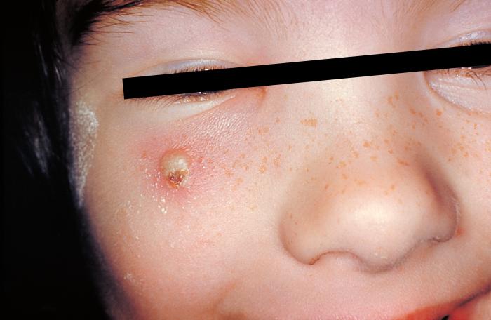
Girl with a secondary skin infection due to chickenpox.
-
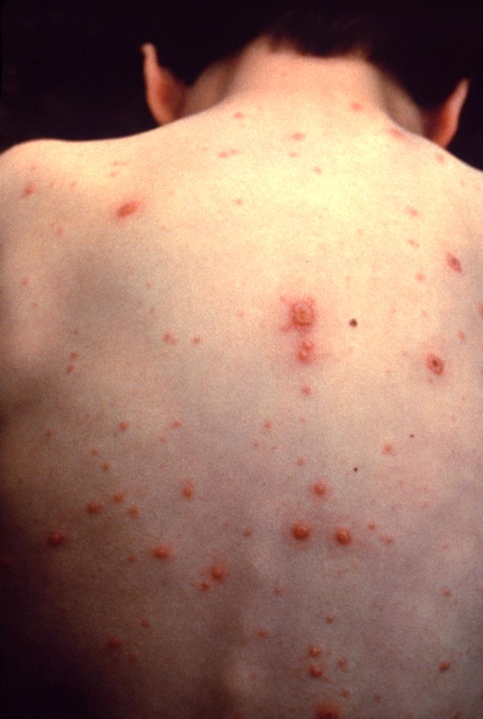
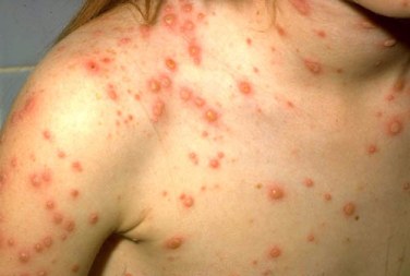
Chickenpox in an unvaccinated child.
-
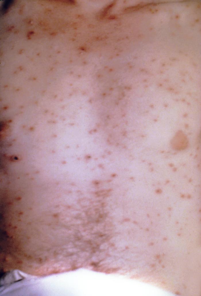
Chickenpox in an unvaccinated adult.
-
Chickenpox in unvaccinated adult.
-
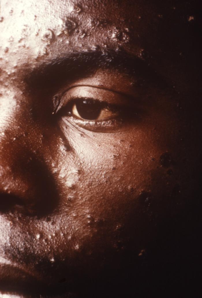
Chickenpox in unvaccinated child.


Vaccinated Individuals
-
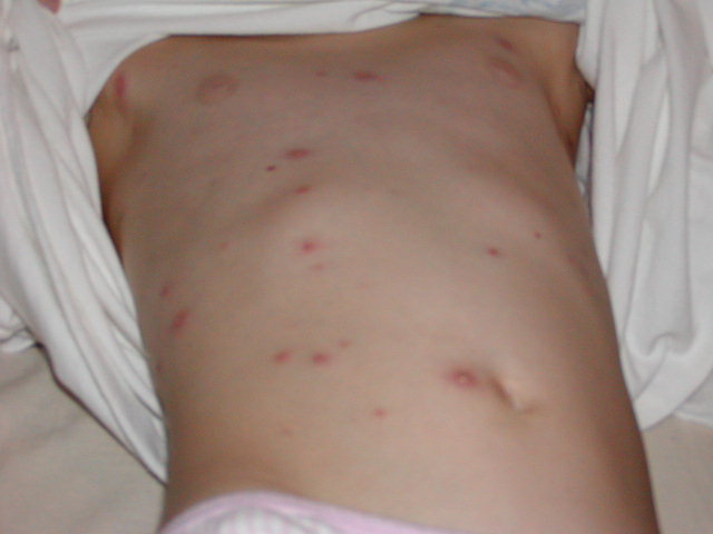
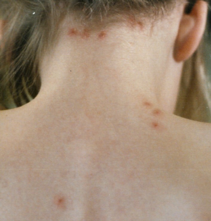
Image of Breakthrough Chickenpox: Back of child with breakthrough varicella.
-
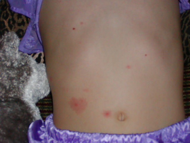
Image of Breakthrough Chickenpox: Back of child with breakthrough varicella.
-
The skin lesions of breakthrough varicella can be macular rather than vesicular. They are rarely bullous or hemorrhagic, and residual scarring is less common.
-
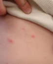
Breakthrough varicella on the abdomen of a vaccinated child.
-
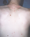
Breakthrough varicella on the back of a vaccinated child.


References
- ↑ McPherson & Pincus: Henry's Clinical Diagnosis and Management by Laboratory Methods, 21st ed., 2007, Chapter 54.
- ↑ 2.00 2.01 2.02 2.03 2.04 2.05 2.06 2.07 2.08 2.09 2.10 2.11 2.12 2.13 2.14 2.15 2.16 2.17 2.18 2.19 2.20 2.21 2.22 2.23 2.24 2.25 2.26 2.27 2.28 2.29 2.30 2.31 2.32 2.33 2.34 2.35 2.36 2.37 "Public Health Image Library (PHIL)".