Bicuspid aortic stenosis chest x ray: Difference between revisions
No edit summary |
Usama Talib (talk | contribs) No edit summary |
||
| (One intermediate revision by one other user not shown) | |||
| Line 1: | Line 1: | ||
{{Bicuspid aortic stenosis}} | {{Bicuspid aortic stenosis}} | ||
{{CMG}}; {{AOEIC}} {{VK}} | {{CMG}}; {{AOEIC}} {{VK}}; {{USAMA}} | ||
==Overview== | ==Overview== | ||
Chest x ray may be used as a diagnostic tool in the evaluation of aortic stenosis. Findings associated with aortic stenosis include [[left ventricular hypertrophy]]. | Chest x ray may be used as a diagnostic tool in the evaluation of aortic stenosis. Findings associated with aortic stenosis include [[left ventricular hypertrophy]]. | ||
| Line 16: | Line 17: | ||
==References== | ==References== | ||
{{reflist|2}} | {{reflist|2}} | ||
{{WS}} | |||
{{WH}} | |||
[[CME Category::Cardiology]] | |||
[[Category:Cardiology]] | [[Category:Cardiology]] | ||
[[Category:Disease]] | [[Category:Disease]] | ||
[[Category:Radiology]] | [[Category:Radiology]] | ||
Latest revision as of 15:54, 5 January 2017
|
Bicuspid aortic stenosis Microchapters |
|
Diagnosis |
|---|
|
Treatment |
|
Bicuspid aortic stenosis chest x ray On the Web |
|
American Roentgen Ray Society Images of Bicuspid aortic stenosis chest x ray |
|
Risk calculators and risk factors for Bicuspid aortic stenosis chest x ray |
Editor-In-Chief: C. Michael Gibson, M.S., M.D. [1]; Associate Editor(s)-In-Chief: Varun Kumar, M.B.B.S. [2]; Usama Talib, BSc, MD [3]
Overview
Chest x ray may be used as a diagnostic tool in the evaluation of aortic stenosis. Findings associated with aortic stenosis include left ventricular hypertrophy.
Chest X Ray

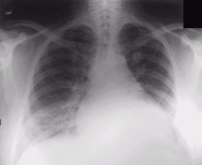
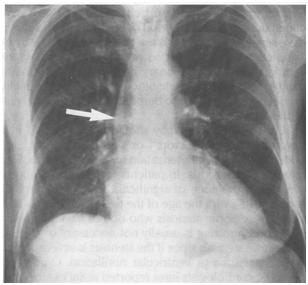
Chest X-ray may show hypertrophied left ventricle if there is aortic stenosis as shown here. In later stages of disease; the left ventricle dilates and the patient may have pulmonary congestion which may be appearant on X-ray. In case of severe aortic stenosis for a long time; the left atrium, pulmonary artery, and right side of heart may become enlarged too.
