Atrial flutter
Template:DiseaseDisorder infobox
|
WikiDoc Resources for Atrial flutter |
|
Articles |
|---|
|
Most recent articles on Atrial flutter Most cited articles on Atrial flutter |
|
Media |
|
Powerpoint slides on Atrial flutter |
|
Evidence Based Medicine |
|
Clinical Trials |
|
Ongoing Trials on Atrial flutter at Clinical Trials.gov Trial results on Atrial flutter Clinical Trials on Atrial flutter at Google
|
|
Guidelines / Policies / Govt |
|
US National Guidelines Clearinghouse on Atrial flutter NICE Guidance on Atrial flutter
|
|
Books |
|
News |
|
Commentary |
|
Definitions |
|
Patient Resources / Community |
|
Patient resources on Atrial flutter Discussion groups on Atrial flutter Patient Handouts on Atrial flutter Directions to Hospitals Treating Atrial flutter Risk calculators and risk factors for Atrial flutter
|
|
Healthcare Provider Resources |
|
Causes & Risk Factors for Atrial flutter |
|
Continuing Medical Education (CME) |
|
International |
|
|
|
Business |
|
Experimental / Informatics |
| Cardiology Network |
 Discuss Atrial flutter further in the WikiDoc Cardiology Network |
| Adult Congenital |
|---|
| Biomarkers |
| Cardiac Rehabilitation |
| Congestive Heart Failure |
| CT Angiography |
| Echocardiography |
| Electrophysiology |
| Cardiology General |
| Genetics |
| Health Economics |
| Hypertension |
| Interventional Cardiology |
| MRI |
| Nuclear Cardiology |
| Peripheral Arterial Disease |
| Prevention |
| Public Policy |
| Pulmonary Embolism |
| Stable Angina |
| Valvular Heart Disease |
| Vascular Medicine |
Editor-In-Chief: C. Michael Gibson, M.S., M.D. [1]
Associate Editor-In-Chief: Cafer Zorkun, M.D., Ph.D. [2]
Please Take Over This Page and Apply to be Editor-In-Chief for this topic: There can be one or more than one Editor-In-Chief. You may also apply to be an Associate Editor-In-Chief of one of the subtopics below. Please mail us [3] to indicate your interest in serving either as an Editor-In-Chief of the entire topic or as an Associate Editor-In-Chief for a subtopic. Please be sure to attach your CV and or biographical sketch.
Overview
Atrial flutter is an abnormal heart rhythm that occurs in the atria of the heart. When it first occurs, it is usually associated with a fast heart rate or tachycardia, and falls into the category of supra-ventricular tachycardias. While this rhythm occurs most often in individuals with cardiovascular disease (eg: hypertension, coronary artery disease, and cardiomyopathy), it may occur spontaneously in people with otherwise normal hearts. It is typically not a stable rhythm, and frequently degenerates into atrial fibrillation. However, it does rarely persist for months to years.
Symptoms
While atrial flutter can sometimes go unnoticed, its onset is often marked by characteristic sensations of regular palpitations. Such sensations usually last until the episode resolves, or until the heart rate is controlled.
Atrial flutter is usually well tolerated initially (fast heart beat is for most people, just a normal response to exercise), however, people with other underlying heart disease or poor exercise tolerance may rapidly develop symptoms, which can include shortness of breath, chest pains, lightheadedness or dizziness, nausea and, in some patients, nervousness and feelings of impending doom.
Prolonged fast flutter may lead to decompensation with loss of normal heart function (heart failure). This may manifest as effort intolerance (exertional breathlessness), nocturnal breathlessness, or swelling of the legs or abdomen.
Pathophysiology: mechanism of action
Atrial flutter is caused by a reentrant rhythm in either the right or left atrium. Typically initiated by a premature electrical impulse arising in the atria, atrial flutter is propogated due to differences in refractory periods of atrial tissue. This creates a self perpetuating loop of electrical activity moving around the atrium.
The impact and symptoms of atrial flutter depend on the heart rate of the patient. Heart rate is a measure of the ventricular rather than atrial activity. Impulses from the atria are conducted to the ventricles through the atrio-ventricular node. Due primarily to its longer refractory period, the AV node exerts a protective effect on heart rate by blocking atrial impulses in excess of about 180 beats/minute (This block is dependent on the age of the patient, and can be calculated roughly by subtracting patient age from 220). If the flutter rate is 300/minute only half of these impulses will be conducted, giving a ventricular rate of 150/minute, ie. 2:1 block. The addition of rate-controlling drugs or conduction system disease can increase this block substantially (see image below).
There are two types of atrial flutter, the common type I and rarer type II.1 Most individuals with atrial flutter will manifest only one of these. Rarely someone may manifest both types; however, they can only manifest one type at a time.
Type I atrial flutter, also known as common atrial flutter or typical atrial flutter, has an atrial rate of 240 to 350 beats/minute. However, this rate may be slowed by antiarrhythmic agents.
The reentrant loop circles the right atrium, passing through the isthmus - a body of fibrous tissue in the lower atrium between the inferior vena cava, and the tricuspid valve. Type I flutter is further divided into two subtypes, known as counterclockwise atrial flutter and clockwise atrial flutter depending on the direction of current passing through the loop. Counterclockwise atrial flutter (known as cephalad-directed atrial flutter) is more commonly seen. The flutter waves in this rhythm are inverted in ecg leads II, III, and aVF. The re-entry loop cycles in the opposite direction in clockwise atrial flutter, thus the flutter waves are upright in II, III, and aVF.
Catheter ablation of the isthmus is a procedure usually available in the electrophysiology laboratory. Eliminating conduction through the isthmus prevents reentry, and if successful, prevents the recurrence of the atrial flutter.
Type II flutter follows a significantly different re-entry pathway to type I flutter, and is typically faster, usually 340–430 beats/minute.
Complications
Although often regarded as a relatively benign rhythm problem, atrial flutter shares the same complications as the related condition atrial fibrillation. There is paucity of published data directly comparing the two, but overall mortality in these conditions appears to be very similar3.
Rate Related
Rapid heart rates may produce significant symptoms in patients with pre-existing heart disease. Even in patients whose hearts are normal to start with, prolonged tachycardia tends to produce ventricular decompensation and heart failure.
Clot formation
Because there is little if any effective contraction of the atria there is stasis (pooling) of blood in the atria. Stasis of blood in susceptible individuals can lead to formation of thrombus (blood clots) within the heart. Thrombus is most likely to form in the atrial appendages. Clot in the left atrial appendage is particularly important since the left side of the heart supplies blood to the entire body. Thus, any thrombus material that dislodges from the this side of the heart can embolize to the brain, with the potentially devastating consequence of a stroke. Thrombus material can of course embolize to any other portion of the body, though usually with a less severe outcome.
Sudden cardiac death
Sudden death is not directly associated with atrial flutter. However, in individuals with a pre-existing accessory conduction pathway, such as the bundle of Kent in Wolff-Parkinson-White syndrome, the accessory pathway may conduct activity from the atria to the ventricles at a rate that the AV node would usually block. Bypassing the AV node, the atrial rate of 300 beats/minute leads to a ventricular rate of 300 beats/minute (1:1 conduction). Even if the ventricles are able to sustain a cardiac output at such a high rates, 1:1 flutter with time may degenerate into ventricular fibrillation, causing hemodynamic collapse and death.
Treatment
In general, atrial flutter should be treated the same as atrial fibrillation. Because both rhythms can lead to the formation of thrombus in the atria, individuals with atrial flutter usually require some form of anticoagulation or anti-platelet agent. Both rhythms can be associated with dangerously fast heart rate and thus require medication for rate and or rhythm control. Additionally, there are some specific considerations particular to treatment of atrial flutter.
Cardioversion
Atrial flutter is considerably more sensitive to electrical direct-current cardioversion than atrial fibrillation, and usually requires a lower energy shock. Conversely, it is relatively resistant to chemical cardioversion, and often deteriorates into atrial fibrillation prior to spontaneous return to sinus rhythm.
Ablation
Because of the reentrant nature of atrial flutter, it is often possible to ablate the circuit that causes atrial flutter. This is done in the electrophysiology lab by causing a ridge of scar tissue that crosses the path of the circuit that causes atrial flutter. Ablation of the isthmus, as discussed above, is a common treatment for typical atrial flutter.
Measurement of Successful Ablation

- Corridor of Widely split double potentials 90-110 ms
- Transisthmus Conduction Intervals
- Counter Clockwise defined as interval between stimulus on lateral wall and proximal coronary sinus electrode.
- Clockwise defined as interval between stimulus in proximal CS and electrodes lateral to line of block.
- Interval measured at 500, 400, and 300 ms. If this value increased by 50% or more this was defined as successs or 150ms
- Pacing at multiple sites. AD>BD and DA>CA
- Bipolar electrograms lateral to line and pace from Proximal CS. Transition of polarity from positive to negative
- 3 pacing site protocol: Pace at two sites lateral (L1R and L2R) to the line on block and on the septal site (S) of the line. Measure the conduction delay from the pacing site to the R wave on the QRS (L1 to R, L2 to R and S to R). If (L1R-L2R) > 0 and (L1R-SR) > 94 then there is a 100% sensitivity and 98% specificity.
Electrocardiographic Findings
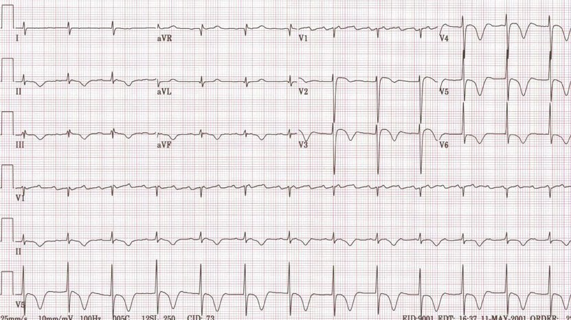
- There are rapid regular undulations (F waves) that cause a sawtooth appearance.
- Best seen in 2,3,F and V1.
- Usually inverted in the inferior leads.
- No isoelectric baselines between the F waves.
- Atrial rate is 250 to 350 Beats Per Minute (BPM).
- Can be faster in infants and children.
- Massive dilation of the atria can lead to a rate < 200 BPM.
- Quinidine can reduce the atrial rate.
- There is a variable ventricular rate depending on the AV conduction.
- The Most common response is 2:1
- 3:1 is uncommon
- 4:1 suggests the existence of an AV conduction defect
- May be associated with complete AV block in which case the RR intervals are regular and the F waves have no constant relationship to the QRS. The ventricular response is usually slow.
- 1:1 conduction may be precipitated by excitement, exercise, induction of anesthesia or any increase in sympathetic tone. It may occur in WPW where the impulses are conducted antegrade through the bypass tract. All these are an emergency.
- During treatment with quinidine the atrial rate may slow sufficiently to permit 1:1 conduction.
- Vagal maneuvers increase the degree of AV block.
- QRS either normal or aberrant depending on preexisting IVCD or aberrant ventricular conduction.
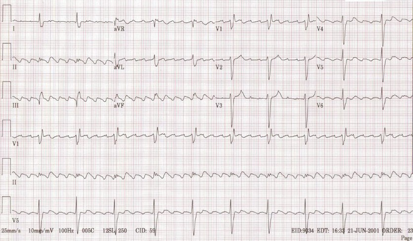
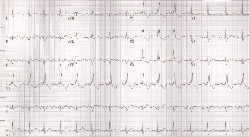
Examples
-
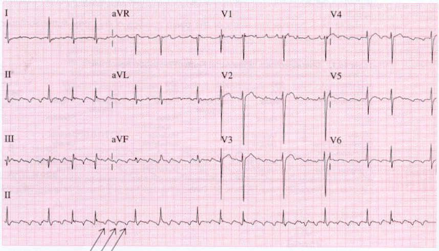
Atrial flutter
-
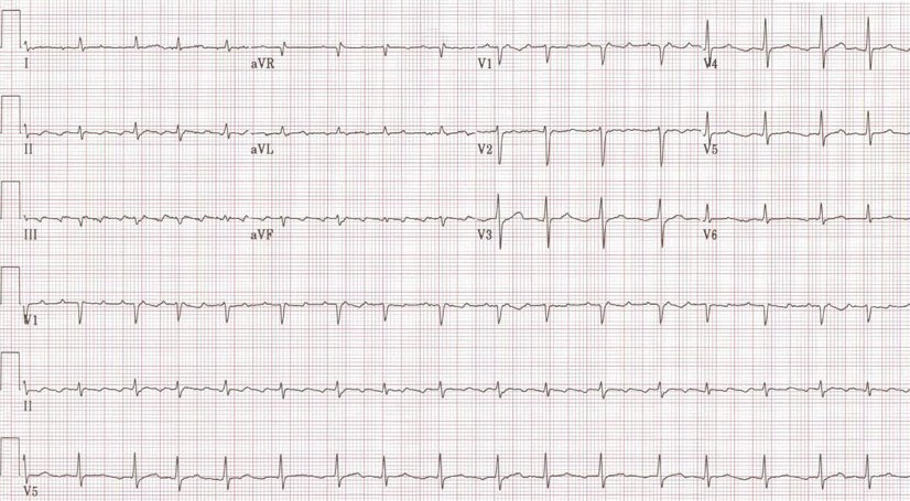
Atrial flutter with variable conduction
-
A very rare condition 1:1 Atrial flutter
-
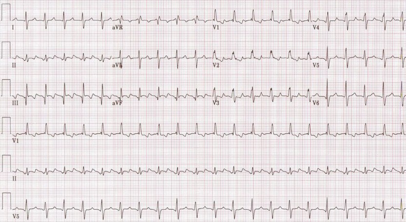
2:1 Atrial flutter
-
3:1 Atrial flutter
-
4:1 Atrial flutter
-
Atrial flutter with RBBB
-

Typical atrial flutter pattern


References
- Chou's Electrocardiography in Clinical Practice, Fifth Edition, Surawicz & Knilans, ISBN 0-7216-8697-4
- Electrophysiologic Testing, Richard N. Fogoros, Blackwell Science, ISBN 0-632-04325-3
- Vidaillet, Granada, et.al. A Population-Based Study of Mortality among Patients with Atrial Fibrillation or Flutter The American Journal of Medicine 113:365-70
External Sources
External Links
See also