Cardiac tamponade electrocardiogram
|
Cardiac tamponade Microchapters |
|
Diagnosis |
|---|
|
Treatment |
|
Case Studies |
|
Cardiac tamponade electrocardiogram On the Web |
|
American Roentgen Ray Society Images of Cardiac tamponade electrocardiogram |
|
Risk calculators and risk factors for Cardiac tamponade electrocardiogram |
Editor-In-Chief: C. Michael Gibson, M.S., M.D. [1]; Associate Editor(s)-in-Chief: Cafer Zorkun, M.D., Ph.D. [2]; Varun Kumar, M.B.B.S.
Overview
The electrocardiogram in cardiac tamponade usually demonstrates sinus tachycardia, and may sometimes show reduced QRS voltage and electrical alternans.
Electrocardiogram
The EKG findings of cardiac tamponade include:[1][2].
Rate
Sinus tachycardia is usually present.
Electrical Alternans
Electrical alternans may be present. When the word alternans is used, the underlying pathophysiology that is most often thought of is alternans due to motion of the heart and its shifting position in relationship to the surface electrodes. The pathophysiologic mechanism underlying the alternation in the height or amplitude of the QRS complex is the swinging or shifting or the electrical axis of the heart. It should be noted that there can also be P wave and T wave alternans attributable to the motion of the heart.
While electrical alternans is frequently thought of in association with pericardial effusion, it should be noted that not all pericardial effusions cause electrical alternans, and that total electrical alternans (involving the p wave, QRS complex and the T wave) is present in just 5-10% of cases of cardiac tamponade.
Low Voltage of the QRS Complex
Low voltage QRS complexes (Low QRS voltage is defined as maximum QRS amplitude in precordial lead < 1 mV and < 0.5 mV in the limb leads due to insulating properties of fluid) [3].
Findings of Pericarditis or Myopericarditis
EKG findings of pericarditis and pericardial effusion may be seen if these conditions are accompanying tamponade. Likewise, ST segment elevation may be present if myopericarditis is present [4].
Electrocardiographic Examples
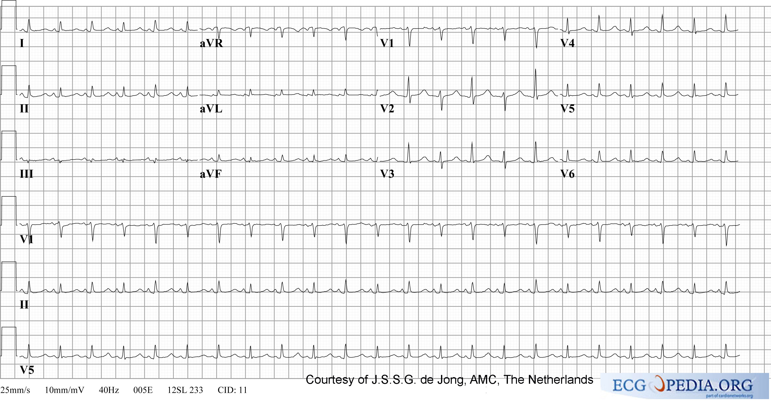
The EKG below shows cardiac tamponade with sinus tachycardia, low voltage QRS complexes and electrical alternans of the QRS complex:

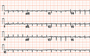
The EKG below shows cardiac tamponade with sinus tachycardia, low voltage QRS complexes and electrical alternans of the QRS complex:

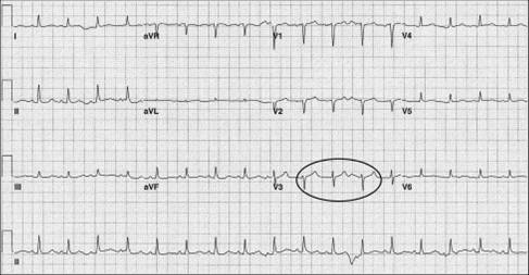
The EKG below shows cardiac tamponade with sinus tachycardia, low voltage QRS complexes and electrical alternans of the QRS complex:

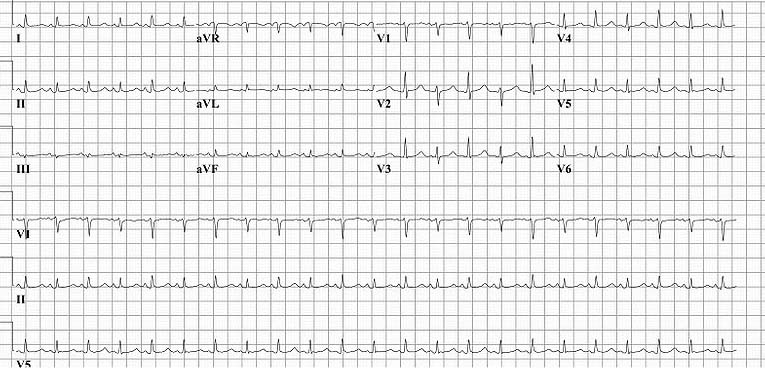
The EKG below shows cardiac tamponade with sinus tachycardia, low voltage QRS complexes and electrical alternans of the QRS complex:

References
- ↑ Jeilan M, Habib A, Colver H, Kundu S, Mohammed N, Loke I (July 2010). "Striking electrical and mechanical alternans associated with cardiac tamponade". BMJ Case Rep. 2010. doi:10.1136/bcr.06.2008.0100. PMC 5497715. PMID 22752620.
- ↑ Dolan, B., Holt, L. (2000). Accident & Emergency: Theory into practice. London: Bailliere Tindall ISBN 978-0702022395
- ↑ Longmore, M., Wilkinson, I.B., Rajagopalan, S. (2004) (6th Ed.). Oxford Handbook of Clinical Medicine. Oxford: Oxford University Press ISBN 9780198568377
- ↑ Dolan, B., Holt, L. (2000). Accident & Emergency: Theory into practice. London: Bailliere Tindall ISBN 978-0702022395