Sandbox:Sahar: Difference between revisions

Jump to navigation Jump to search
No edit summary
No edit summary
Line 12: Line 12:
! style="background: #4479BA; width: 150px;" | {{fontcolor|#FFF| Microscopic Appearance}}
|-
|-
! style="padding: 5px 5px; background: #DCDCDC; " align="left" |Papillary Thyroid Cancer<ref name="FaginMitsiades2008">{{cite journal|last1=Fagin|first1=James A.|last2=Mitsiades|first2=Nicholas|title=Molecular pathology of thyroid cancer: diagnostic and clinical implications|journal=Best Practice & Research Clinical Endocrinology & Metabolism|volume=22|issue=6|year=2008|pages=955–969|issn=1521690X|doi=10.1016/j.beem.2008.09.017}}</ref><ref name="Schlumberger1998">{{cite journal|last1=Schlumberger|first1=Martin Jean|title=Papillary and Follicular Thyroid Carcinoma|journal=New England Journal of Medicine|volume=338|issue=5|year=1998|pages=297–306|issn=0028-4793|doi=10.1056/NEJM199801293380506}}</ref><ref name="pmid20001718">{{cite journal |vauthors=Sipos JA |title=Advances in ultrasound for the diagnosis and management of thyroid cancer |journal=Thyroid |volume=19 |issue=12 |pages=1363–72 |date=December 2009 |pmid=20001718 |doi=10.1089/thy.2009.1608 |url=}}</ref>
! style="padding: 5px 5px; background: #DCDCDC; " align="left" |
| style="padding: 5px 5px; background: #F5F5F5;" align="left" |
| style="padding: 5px 5px; background: #F5F5F5;" align="left" |
* More common in the middle aged (30 - 50 years of age)
Line 47: Line 47:
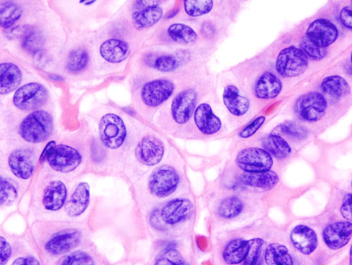
| style="padding: 5px 5px; background: #F5F5F5;" align="left" |[[Image:Thyroid papillary carcinoma histopathology (3).jpg|thumb|none|200px|Source:Wikimedia commons ]]
| style="padding: 5px 5px; background: #F5F5F5;" align="left" |[[Image:Thyroid papillary carcinoma histopathology (3).jpg|thumb|none|200px|Source:Wikimedia commons ]]
|-
|-
! style="padding: 5px 5px; background: #DCDCDC; " align="left" |Follicular Thyroid Cancer<ref name="Schlumberger1998">{{cite journal|last1=Schlumberger|first1=Martin Jean|title=Papillary and Follicular Thyroid Carcinoma|journal=New England Journal of Medicine|volume=338|issue=5|year=1998|pages=297–306|issn=0028-4793|doi=10.1056/NEJM199801293380506}}</ref><ref name="pmid20001718">{{cite journal |vauthors=Sipos JA |title=Advances in ultrasound for the diagnosis and management of thyroid cancer |journal=Thyroid |volume=19 |issue=12 |pages=1363–72 |date=December 2009 |pmid=20001718 |doi=10.1089/thy.2009.1608 |url=}}</ref><ref name="pmid2019455">{{cite journal |vauthors=Pettersson B, Adami HO, Wilander E, Coleman MP |title=Trends in thyroid cancer incidence in Sweden, 1958-1981, by histopathologic type |journal=Int. J. Cancer |volume=48 |issue=1 |pages=28–33 |date=April 1991 |pmid=2019455 |doi=10.1002/ijc.2910480106 |url=}}</ref>
! style="padding: 5px 5px; background: #DCDCDC; " align="left" |
| style="padding: 5px 5px; background: #F5F5F5;" align="left" |
| style="padding: 5px 5px; background: #F5F5F5;" align="left" |
* Peak [[incidence]] at 40 - 60 years of age
Line 80: Line 80:
| style="padding: 5px 5px; background: #F5F5F5;" align="left" |[[Image:Metastatic follicular thyroid carcinoma - Case 264.jpg|thumb|none|200px|Source:Wikimedia common ]]
| style="padding: 5px 5px; background: #F5F5F5;" align="left" |[[Image:Metastatic follicular thyroid carcinoma - Case 264.jpg|thumb|none|200px|Source:Wikimedia common ]]
|-
|-
! style="padding: 5px 5px; background: #DCDCDC;" align="left" |Medullary Thyroid Cancer<ref name="pmid6690009">{{cite journal |vauthors=Busnardo B, Girelli ME, Simioni N, Nacamulli D, Busetto E |title=Nonparallel patterns of calcitonin and carcinoembryonic antigen levels in the follow-up of medullary thyroid carcinoma |journal=Cancer |volume=53 |issue=2 |pages=278–85 |date=January 1984 |pmid=6690009 |doi=10.1002/1097-0142(19840115)53:2<278::aid-cncr2820530216>3.0.co;2-z |url=}}</ref><ref name="pmid10699905">{{cite journal |vauthors=Kebebew E, Ituarte PH, Siperstein AE, Duh QY, Clark OH |title=Medullary thyroid carcinoma: clinical characteristics, treatment, prognostic factors, and a comparison of staging systems |journal=Cancer |volume=88 |issue=5 |pages=1139–48 |date=March 2000 |pmid=10699905 |doi=10.1002/(sici)1097-0142(20000301)88:5<1139::aid-cncr26>3.0.co;2-z |url=}}</ref><ref name="HofstraLandsvater1994">{{cite journal|last1=Hofstra|first1=Robert M. W.|last2=Landsvater|first2=Rudy M.|last3=Ceccherini|first3=Isabella|last4=Stulp|first4=Rein P.|last5=Stelwagen|first5=Tineke|last6=Luo|first6=Yin|last7=Pasini|first7=Barbara|last8=Hoppener|first8=Jo W. M.|last9=van Amstel|first9=Hans Kristian Ploos|last10=Romeo|first10=Giovanni|last11=Lips|first11=Cornells J. M.|last12=Buys|first12=Charles H. C. M.|title=A mutation in the RET proto-oncogene associated with multiple endocrine neoplasia type 2B and sporadic medullary thyroid carcinoma|journal=Nature|volume=367|issue=6461|year=1994|pages=375–376|issn=0028-0836|doi=10.1038/367375a0}}</ref><ref name="pmid20001718">{{cite journal |vauthors=Sipos JA |title=Advances in ultrasound for the diagnosis and management of thyroid cancer |journal=Thyroid |volume=19 |issue=12 |pages=1363–72 |date=December 2009 |pmid=20001718 |doi=10.1089/thy.2009.1608 |url=}}</ref>
! style="padding: 5px 5px; background: #DCDCDC;" align="left" |Medullary Thyroid Cancer<ref name="pmid20001718">{{cite journal |vauthors=Sipos JA |title=Advances in ultrasound for the diagnosis and management of thyroid cancer |journal=Thyroid |volume=19 |issue=12 |pages=1363–72 |date=December 2009 |pmid=20001718 |doi=10.1089/thy.2009.1608 |url=}}</ref>


| style="padding: 5px 5px; background: #F5F5F5;" align="left" |
| style="padding: 5px 5px; background: #F5F5F5;" align="left" |
Line 111: Line 111:
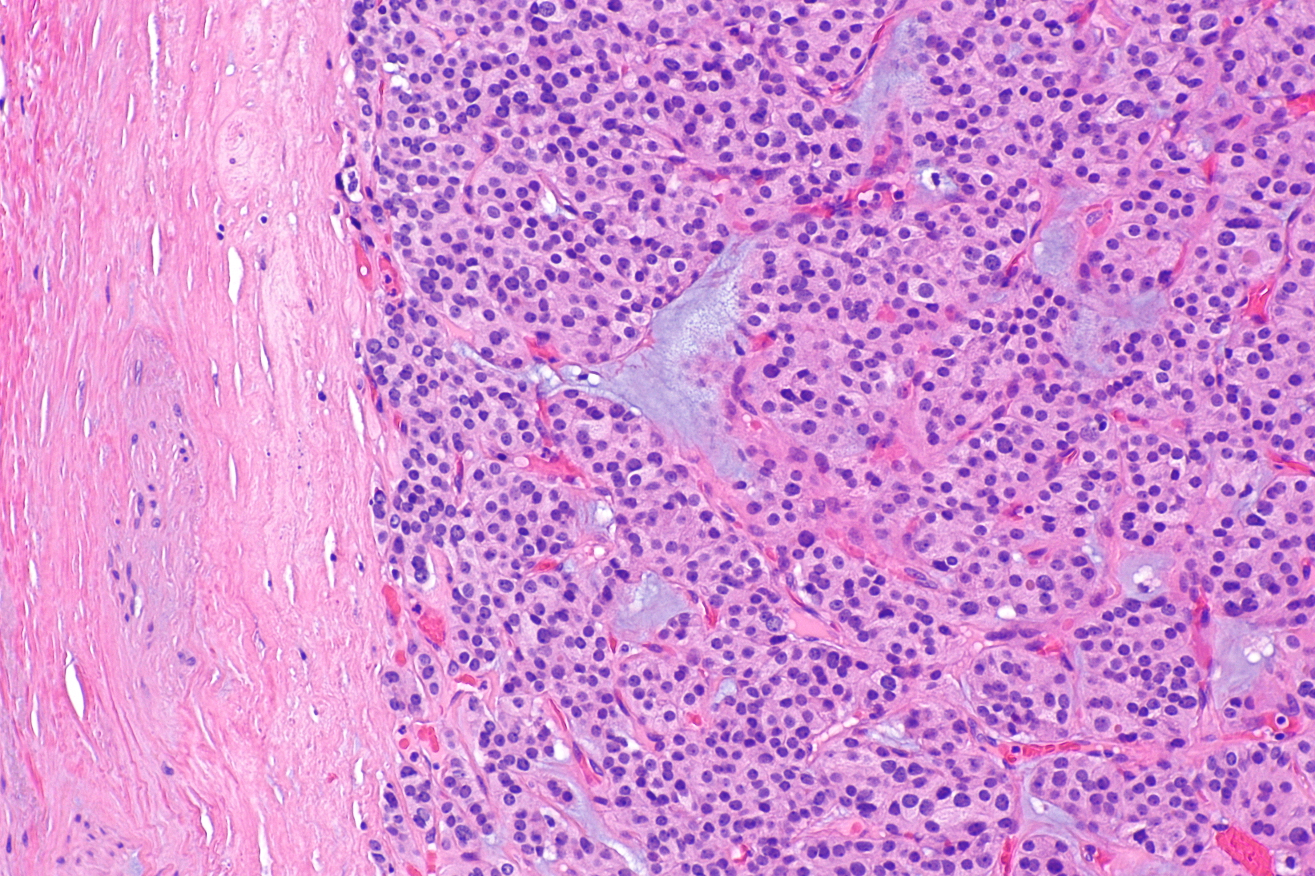
| style="padding: 5px 5px; background: #F5F5F5;" align="left" |[[File:Thyroid MedullaryCarcinoma SpindleCell LP PA.JPG|thumb|none|200px|Source:Wikimedia common ]]
| style="padding: 5px 5px; background: #F5F5F5;" align="left" |[[File:Thyroid MedullaryCarcinoma SpindleCell LP PA.JPG|thumb|none|200px|Source:Wikimedia common ]]
|-
|-
! style="padding: 5px 5px; background: #DCDCDC;" align="left" |Anaplastic Thyroid Cancer<ref name="pmid21772843">{{cite journal |vauthors=Nagaiah G, Hossain A, Mooney CJ, Parmentier J, Remick SC |title=Anaplastic thyroid cancer: a review of epidemiology, pathogenesis, and treatment |journal=J Oncol |volume=2011 |issue= |pages=542358 |date=2011 |pmid=21772843 |pmc=3136148 |doi=10.1155/2011/542358 |url=}}</ref><ref name="pmid2794956">{{cite journal |vauthors=Chang TC, Liaw KY, Kuo SH, Chang CC, Chen FW |title=Anaplastic thyroid carcinoma: review of 24 cases, with emphasis on cytodiagnosis and leukocytosis |journal=Taiwan Yi Xue Hui Za Zhi |volume=88 |issue=6 |pages=551–6 |date=June 1989 |pmid=2794956 |doi= |url=}}</ref><ref name="pmid1695118">{{cite journal |vauthors=Venkatesh YS, Ordonez NG, Schultz PN, Hickey RC, Goepfert H, Samaan NA |title=Anaplastic carcinoma of the thyroid. A clinicopathologic study of 121 cases |journal=Cancer |volume=66 |issue=2 |pages=321–30 |date=July 1990 |pmid=1695118 |doi=10.1002/1097-0142(19900715)66:2<321::aid-cncr2820660221>3.0.co;2-a |url=}}</ref>
! style="padding: 5px 5px; background: #DCDCDC;" align="left" |Anaplastic Thyroid Cancer
| style="padding: 5px 5px; background: #F5F5F5;" align="left" |
| style="padding: 5px 5px; background: #F5F5F5;" align="left" |
* More common among older individuals
Line 141: Line 141:
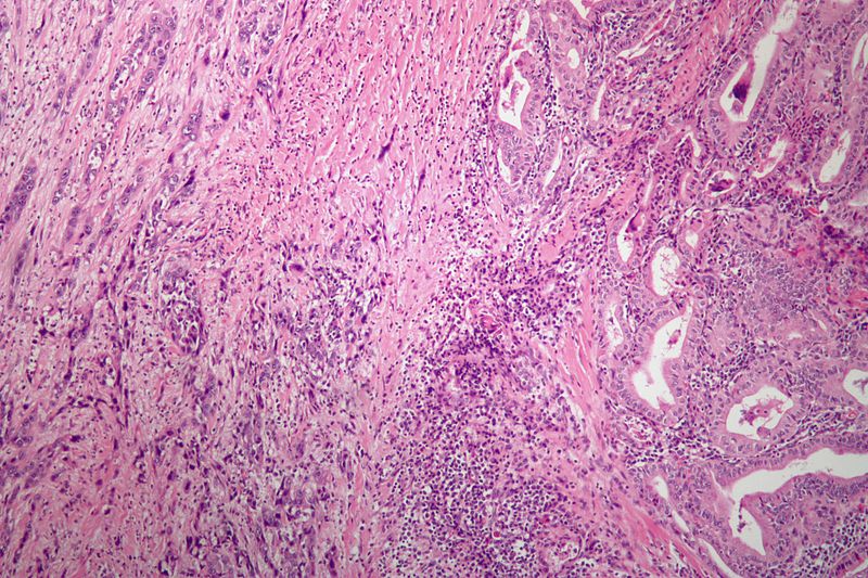
| style="padding: 5px 5px; background: #F5F5F5;" align="left" |[[File:Anaplastic thyroid carcinoma low mag.jpg|thumb|none|200px|Source:Wikimedia common ]]
| style="padding: 5px 5px; background: #F5F5F5;" align="left" |[[File:Anaplastic thyroid carcinoma low mag.jpg|thumb|none|200px|Source:Wikimedia common ]]
|-
|-
! style="padding: 5px 5px; background: #DCDCDC;" align="left" |Follicular Adenoma<ref name="MathurOlson2014">{{cite journal|last1=Mathur|first1=Aarti|last2=Olson|first2=Matthew T.|last3=Zeiger|first3=Martha A.|title=Follicular Lesions of the Thyroid|journal=Surgical Clinics of North America|volume=94|issue=3|year=2014|pages=499–513|issn=00396109|doi=10.1016/j.suc.2014.02.005}}</ref>
! style="padding: 5px 5px; background: #DCDCDC;" align="left" |Follicular Adenoma
| style="padding: 5px 5px; background: #F5F5F5;" align="left" |
| style="padding: 5px 5px; background: #F5F5F5;" align="left" |
* More commonly affects individuals older than 50 years of age
Line 165: Line 165:
| style="padding: 5px 5px; background: #F5F5F5;" align="left" |[[File:Follicular adenoma -- intermed mag.jpg|thumb|none|200px|Source:Wikimedia common ]]
| style="padding: 5px 5px; background: #F5F5F5;" align="left" |[[File:Follicular adenoma -- intermed mag.jpg|thumb|none|200px|Source:Wikimedia common ]]
|-
|-
! style="padding: 5px 5px; background: #DCDCDC;" align="left" |Multinodular Goiter<ref name="pmid8197088">{{cite journal |vauthors=Bronshteĭn ME, Makarov AD, Artemova AM, Bazarova EN, Kozlov GI |title=[Morphology of the thyroid tissue in multinodular euthyroid goiter] |language=Russian |journal=Probl Endokrinol (Mosk) |volume=40 |issue=2 |pages=36–9 |date=1994 |pmid=8197088 |doi= |url=}}</ref>
! style="padding: 5px 5px; background: #DCDCDC;" align="left" |Multinodular Goiter
| style="padding: 5px 5px; background: #F5F5F5;" align="left" |
| style="padding: 5px 5px; background: #F5F5F5;" align="left" |
* Commonly affects individuals older than 60 years of age
Line 189: Line 189:
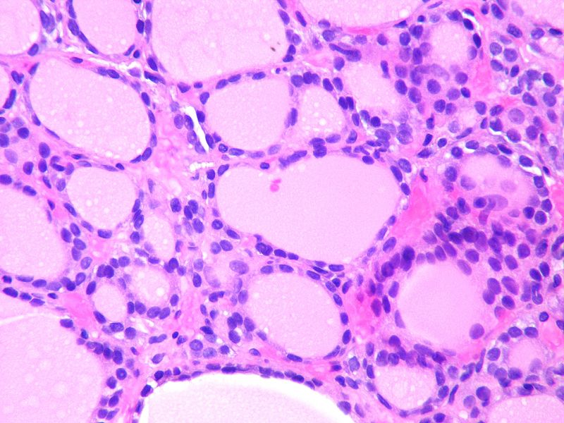
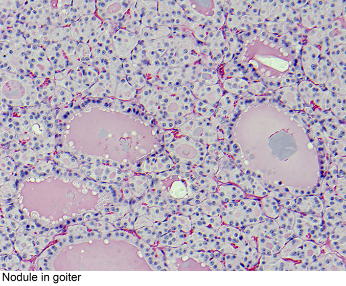
| style="padding: 5px 5px; background: #F5F5F5;" align="left" |[[File:ThyroidnodularSatturwar08.jpg|thumb|none|200px|Source:pathology outline, case courtesy of Dr. Swati Satturwar]]
| style="padding: 5px 5px; background: #F5F5F5;" align="left" |[[File:ThyroidnodularSatturwar08.jpg|thumb|none|200px|Source:pathology outline, case courtesy of Dr. Swati Satturwar]]
|-
|-
! style="padding: 5px 5px; background: #DCDCDC; " align="left" |Thyroid Lymphoma<ref name="pmid8838117">{{cite journal |vauthors=Pedersen RK, Pedersen NT |title=Primary non-Hodgkin's lymphoma of the thyroid gland: a population based study |journal=Histopathology |volume=28 |issue=1 |pages=25–32 |date=January 1996 |pmid=8838117 |doi= |url=}}</ref>
! style="padding: 5px 5px; background: #DCDCDC; " align="left" |Thyroid Lymphoma
<ref name="pmid3141260">{{cite journal |vauthors=Hyjek E, Isaacson PG |title=Primary B cell lymphoma of the thyroid and its relationship to Hashimoto's thyroiditis |journal=Hum. Pathol. |volume=19 |issue=11 |pages=1315–26 |date=November 1988 |pmid=3141260 |doi=10.1016/s0046-8177(88)80287-9 |url=}}</ref><ref name="pmid3759532">{{cite journal |vauthors=Tupchong L, Hughes F, Harmer CL |title=Primary lymphoma of the thyroid: clinical features, prognostic factors, and results of treatment |journal=Int. J. Radiat. Oncol. Biol. Phys. |volume=12 |issue=10 |pages=1813–21 |date=October 1986 |pmid=3759532 |doi=10.1016/0360-3016(86)90324-x |url=}}</ref><ref name="pmid17042683">{{cite journal |vauthors=Ota H, Ito Y, Matsuzuka F, Kuma S, Fukata S, Morita S, Kobayashi K, Nakamura Y, Kakudo K, Amino N, Miyauchi A |title=Usefulness of ultrasonography for diagnosis of malignant lymphoma of the thyroid |journal=Thyroid |volume=16 |issue=10 |pages=983–7 |date=October 2006 |pmid=17042683 |doi=10.1089/thy.2006.16.983 |url=}}</ref>
 
| style="padding: 5px 5px; background: #F5F5F5;" align="left" |
| style="padding: 5px 5px; background: #F5F5F5;" align="left" |
* Affects [[Adult|adults]] or elderly
Line 224: Line 224:
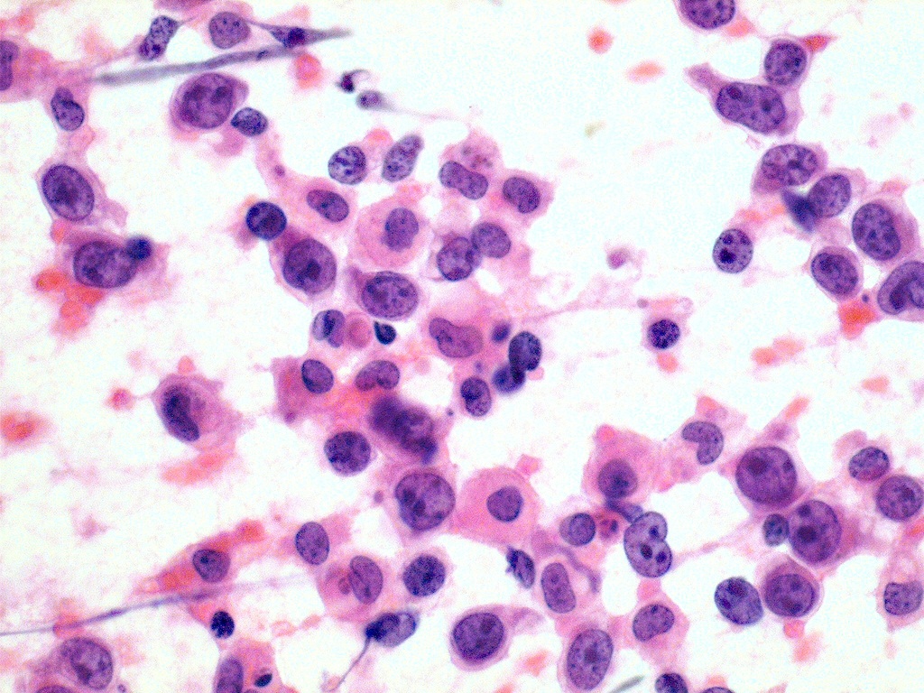
| style="padding: 5px 5px; background: #F5F5F5;" align="left" |[[File:Thyroid lymphoma large cell type fine needle aspiration biop.jpeg|thumb|none|200px|Source:pathology outline, case courtesy of Dr. Mark R. Wick]]
| style="padding: 5px 5px; background: #F5F5F5;" align="left" |[[File:Thyroid lymphoma large cell type fine needle aspiration biop.jpeg|thumb|none|200px|Source:pathology outline, case courtesy of Dr. Mark R. Wick]]
|}
|}
<references />

Revision as of 18:05, 16 January 2020

Disease Name Age of Onset Gender Preponderance Signs/Symptoms Imaging Feature(s) Macroscopic Feature(s) Microscopic Feature(s) Laboratory Findings(s) Other Feature(s) Microscopic Appearance
  • More common in the middle aged (30 - 50 years of age)
  • More commonly affects women
Source:Wikimedia commons
  • More commonly affects women
Source:Wikimedia common
Medullary Thyroid Cancer[1]
  • Incidence increases with age
  • More common in the 3rd to 4th decades of life
  • Both genders are affected equally
  • Single non-encapsulated mass
  • Gray-tan color
Source:Wikimedia common
Anaplastic Thyroid Cancer
  • More common among older individuals
  • Mean age at diagnosis is 65 years
  • More commonly affects women
    • Irregular margin
  • Imaging features are not characteristic of this cancer
Source:Wikimedia common
Follicular Adenoma
  • More commonly affects individuals older than 50 years of age
  • More commonly affects women
  • Solitary nodule which may show echogenicity or not
  • Solitary, spherical, and encapsulated lesion
  • Well demarcated from the surrounding parenchyma
  • Functional adenoma:
    • Elevated T3, T4
    • Decreased TSH
  • May be considered functional or hot
  • May be considered non-functional or cold
Source:Wikimedia common
Multinodular Goiter
  • Commonly affects individuals older than 60 years of age
  • More commonly affects women
Source:pathology outline, case courtesy of Dr. Swati Satturwar
Thyroid Lymphoma
  • More common among women
Source:pathology outline, case courtesy of Dr. Mark R. Wick
  1. Sipos JA (December 2009). "Advances in ultrasound for the diagnosis and management of thyroid cancer". Thyroid. 19 (12): 1363–72. doi:10.1089/thy.2009.1608. PMID 20001718.