Retinoblastoma MRI: Difference between revisions
Jump to navigation
Jump to search
(→MRI) |
No edit summary |
||
| Line 11: | Line 11: | ||
*On T1 image:Hyperintense [[mass]] compared to [[vitreous body]] | *On T1 image:Hyperintense [[mass]] compared to [[vitreous body]] | ||
*On T2 image:Hypointense [[mass]] compared to [[vitreous body]] | *On T2 image:Hypointense [[mass]] compared to [[vitreous body]] | ||
*It should be mentioned that [[MRI]] is accurate for [[tumor]] [[Cancer staging|staging]] and evaluation of [[metastatic]] [[risk factors]].<ref name="de GraafBarkhof2005">{{cite journal|last1=de Graaf|first1=Pim|last2=Barkhof|first2=Frederik|last3=Moll|first3=Annette C.|last4=Imhof|first4=Saskia M.|last5=Knol|first5=Dirk L.|last6=van der Valk|first6=Paul|last7=Castelijns|first7=Jonas A.|title=Retinoblastoma: MR Imaging Parameters in Detection of Tumor Extent|journal=Radiology|volume=235|issue=1|year=2005|pages=197–207|issn=0033-8419|doi=10.1148/radiol.2351031301}}</ref> | *It should be mentioned that [[MRI]] is [[Accuracy|accurate]] for [[tumor]] [[Cancer staging|staging]] and evaluation of [[metastatic]] [[risk factors]].<ref name="de GraafBarkhof2005">{{cite journal|last1=de Graaf|first1=Pim|last2=Barkhof|first2=Frederik|last3=Moll|first3=Annette C.|last4=Imhof|first4=Saskia M.|last5=Knol|first5=Dirk L.|last6=van der Valk|first6=Paul|last7=Castelijns|first7=Jonas A.|title=Retinoblastoma: MR Imaging Parameters in Detection of Tumor Extent|journal=Radiology|volume=235|issue=1|year=2005|pages=197–207|issn=0033-8419|doi=10.1148/radiol.2351031301}}</ref> | ||
*This [[Imaging studies|imaging study]] is capable to discriminate the [[tumor]] with secondary [[retinal detachment]] from other [[causes]] of subretinal effusion or [[Retinal detachment|detachment]], such as coat's [[disease]], primary hyperplastic [[vitreous]] and [[retinopathy of prematurity]]. | *This [[Imaging studies|imaging study]] is capable to discriminate the [[tumor]] with secondary [[retinal detachment]] from other [[causes]] of subretinal effusion or [[Retinal detachment|detachment]], such as coat's [[disease]], primary hyperplastic [[vitreous]] and [[retinopathy of prematurity]]. | ||
*Also, some expert argue that [[CT-scans|CT imaging studies]] in [[bilateral]] [[retinoblastoma]] individuals will increase the risk of individuals for future [[malignancies]]. [[MRI]] is not associated with such risk. | *Also, some expert argue that [[CT-scans|CT imaging studies]] in [[bilateral]] [[retinoblastoma]] individuals will increase the risk of individuals for future [[malignancies]]. [[MRI]] is not associated with such risk. | ||
Revision as of 20:09, 14 May 2019
|
Retinoblastoma Microchapters |
|
Diagnosis |
|---|
|
Treatment |
|
Case Studies |
|
Retinoblastoma MRI On the Web |
|
American Roentgen Ray Society Images of Retinoblastoma MRI |
Editor-In-Chief: C. Michael Gibson, M.S., M.D. [1]; Associate Editor(s)-in-Chief: Sahar Memar Montazerin, M.D.[2] Simrat Sarai, M.D. [3]
Overview
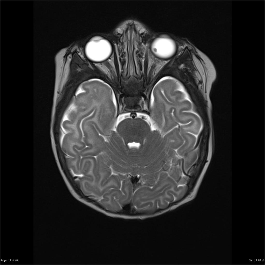
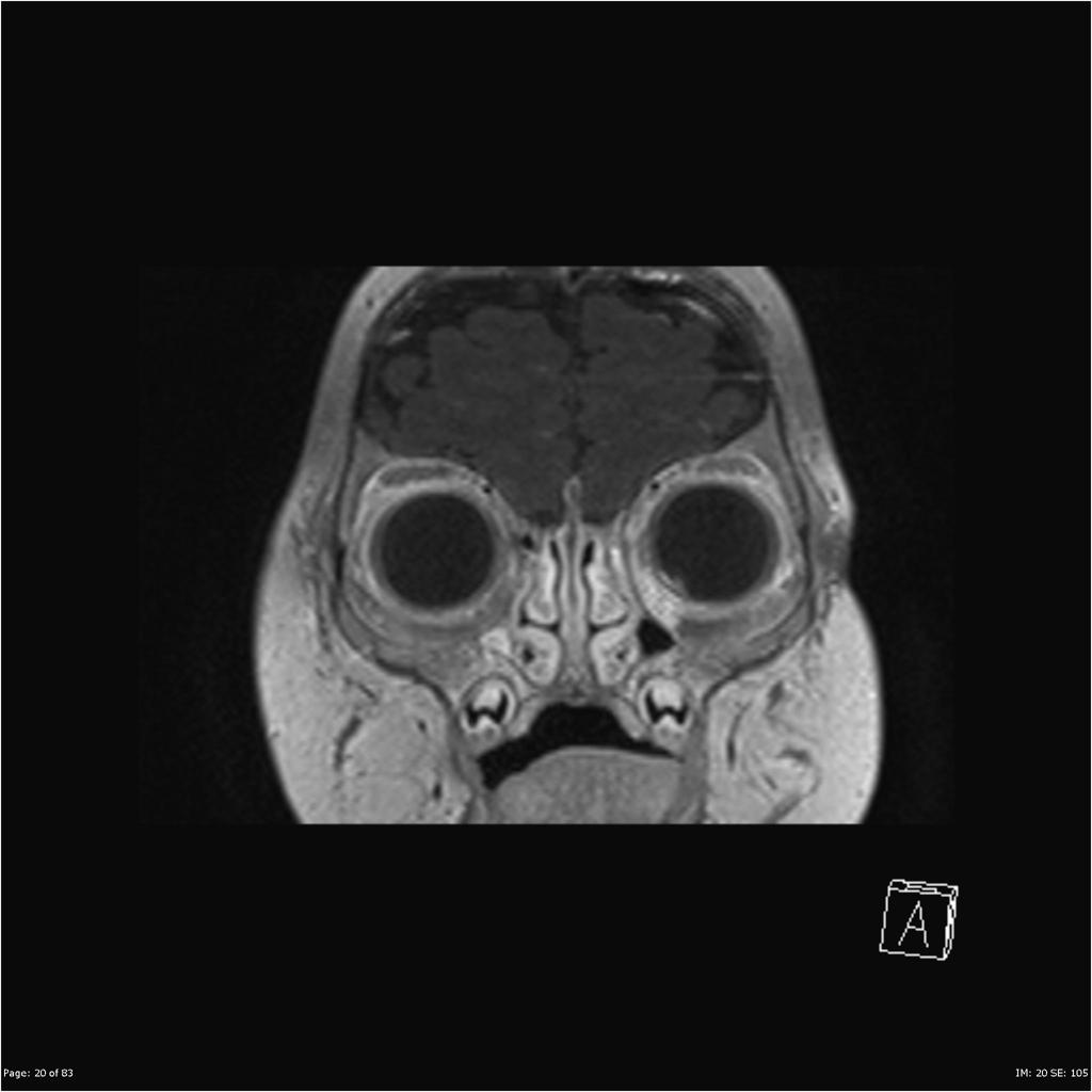
On head and neck MRI, retinoblastoma is characterized by hyperintense mass on T1-weighted MRI and hypointense mass on T2-weighted MRI.
MRI
- MRI imaging study of retinoblastoma first introduced in 1980.[1]
- This method is used as an extra diagnostic method to CT scan in suspected retinoblastoma cases especially in those whose CT scan imaging is doubtful for the presence of extra-ocular extension.[2]
- MRI diagnostic value in the diagnosis of advanced retinoblastoma is greater than CT scan.[3]
MRI findings diagnostic of retinoblastoma are:
- On T1 image:Hyperintense mass compared to vitreous body
- On T2 image:Hypointense mass compared to vitreous body
- It should be mentioned that MRI is accurate for tumor staging and evaluation of metastatic risk factors.[4]
- This imaging study is capable to discriminate the tumor with secondary retinal detachment from other causes of subretinal effusion or detachment, such as coat's disease, primary hyperplastic vitreous and retinopathy of prematurity.
- Also, some expert argue that CT imaging studies in bilateral retinoblastoma individuals will increase the risk of individuals for future malignancies. MRI is not associated with such risk.
- MR imaging is the study of choice to rule out extra-ocular extension of the tumor.[2]

|

|
References
- ↑ Schueler, A O (2003). "High resolution magnetic resonance imaging of retinoblastoma". British Journal of Ophthalmology. 87 (3): 330–335. doi:10.1136/bjo.87.3.330. ISSN 0007-1161.
- ↑ 2.0 2.1 Meel R, Radhakrishnan V, Bakhshi S (April 2012). "Current therapy and recent advances in the management of retinoblastoma". Indian J Med Paediatr Oncol. 33 (2): 80–8. doi:10.4103/0971-5851.99731. PMC 3439795. PMID 22988349.
- ↑ de Jong, Marcus C.; de Graaf, Pim; Noij, Daniel P.; Göricke, Sophia; Maeder, Philippe; Galluzzi, Paolo; Brisse, Hervé J.; Moll, Annette C.; Castelijns, Jonas A. (2014). "Diagnostic Performance of Magnetic Resonance Imaging and Computed Tomography for Advanced Retinoblastoma". Ophthalmology. 121 (5): 1109–1118. doi:10.1016/j.ophtha.2013.11.021. ISSN 0161-6420.
- ↑ de Graaf, Pim; Barkhof, Frederik; Moll, Annette C.; Imhof, Saskia M.; Knol, Dirk L.; van der Valk, Paul; Castelijns, Jonas A. (2005). "Retinoblastoma: MR Imaging Parameters in Detection of Tumor Extent". Radiology. 235 (1): 197–207. doi:10.1148/radiol.2351031301. ISSN 0033-8419.