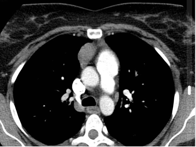
Thymoma CT scan

Jump to navigation Jump to search

Thymoma Microchapters

Home

Patient Information

Overview

Historical Perspective

Classification

Pathophysiology

Causes

Differentiating Thymoma from other Diseases

Epidemiology and Demographics

Risk Factors

Screening

Natural History, Complications and Prognosis

Diagnosis

Diagnostic Study of Choice

History and Symptoms

Physical Examination

Laboratory Findings

Electrocardiogram

X-ray

Echocardiography and Ultrasound

CT scan

MRI

Other Imaging Findings

Other Diagnostic Studies

Treatment

Medical Therapy

Surgery

Primary Prevention

Secondary Prevention

Cost-Effectiveness of Therapy

Future or Investigational Therapies

Case Studies

Case #1

Thymoma CT scan On the Web

Most recent articles

Most cited articles

Review articles

CME Programs

Powerpoint slides

Images

American Roentgen Ray Society Images of Thymoma CT scan

All Images
X-rays
Echo & Ultrasound
CT Images
MRI

Ongoing Trials at Clinical Trials.gov

US National Guidelines Clearinghouse

NICE Guidance

FDA on Thymoma CT scan

CDC on Thymoma CT scan

Thymoma CT scan in the news

Blogs on Thymoma CT scan

Directions to Hospitals Treating Thymoma

Risk calculators and risk factors for Thymoma CT scan

Editor-In-Chief: C. Michael Gibson, M.S., M.D. [4] Associate Editor(s)-in-Chief: Amr Marawan, M.D. [5]Ahmad Al Maradni, M.D. [6]

Overview

Computed Tomography scan may be diagnostic of thymoma. The tumor is generally located inside the thymus, and can be calcified. Increased vascular enhancement can be indicative of malignancy, as can pleural deposits.

CT scan

  • Key CT scan findings in thymoma include:
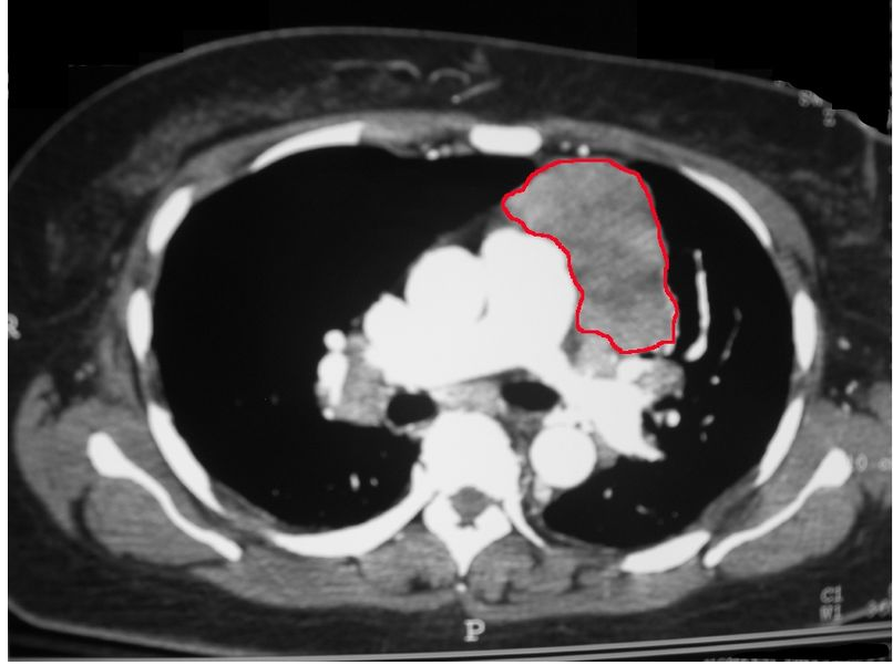
CT scan of the chest revealing a large necrotic mass in the left anterior mediastinum (later proven to be a thymoma) and bilateral hilar lymphadenopathy.
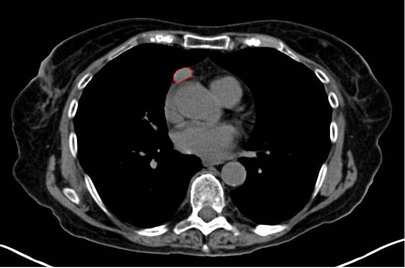
CT shows thymoma (Thymoma, Stage IIA. CT scan, axial).
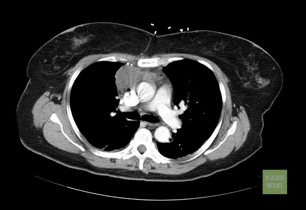
CT shows Thymoma (Image courtesy of RadsWiki and copylefted).
A 43-year-old female with myasthenia gravis. Axial chest CT demonstrates a small soft tissue mass in the anterior mediastinum, proven to be thymoma.[1]
A 43-year-old female with myasthenia gravis. Coronal chest CT demonstrates a small soft tissue mass in the anterior mediastinum, proven to be thymoma.[1]
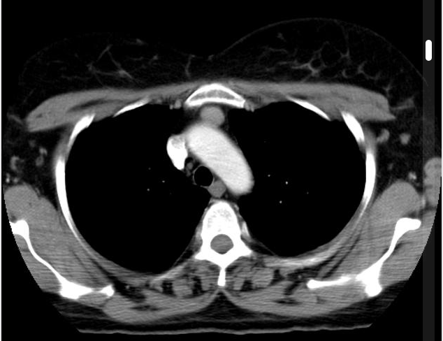
CT shows invasive Thymoma.
CT shows anterior mediastinal mass, proven to be a thymoma.
CT demonstrates a well circumscribed soft tissue mass in the anterior mediastinum, proven to be thymoma.[2]
CT demonstrates a well circumscribed soft tissue mass in the anterior mediastinum, proven to be a thymoma.[2]
CT showing well defined solid lesion is seen in the anterior mediastinum. No calcification cystic or fat component is seen within.[3]

References

  1. 1.0 1.1 Image courtesy of Dr Hani Al Salam. Radiopaedia [1].Creative Commons BY-SA-NC
  2. 2.0 2.1 2.2 Image courtesy of Dr Frank Gaillard. Radiopaedia [2].Creative Commons BY-SA-NC
  3. Image courtesy of Dr Prashant Mudgal. Radiopaedia [3].Creative Commons BY-SA-NC

Template:WikiDoc Sources