Surface epithelial-stromal tumor
Editor-In-Chief: C. Michael Gibson, M.S., M.D. [1] Associate Editor(s)-in-Chief: Maria Fernanda Villarreal, M.D. [2]
Synonyms and keywords: Borderline ovarian tumors; Ovarian epithelial neoplasms
Overview
Surface epithelial-stromal tumors are a class of ovarian neoplasms that may be benign or malignant. Neoplasms in this group are thought to be derived from the ovarian surface epithelium (modified peritoneum) or from ectopic endometrial or Fallopian tube (tubal) tissue. This group of tumors accounts for 90% to 95% of all cases of ovarian cancer.[1][2] The pathogenesis of surface epithelial-stromal tumor is characterized by the overgrowth of the ovarian surface epithelium. Common risk factors in the development of surface epithelial-stromal tumor, include: nulliparity, early menopause, gonadal dysgenesis, family history (e.g. BRCA1/BRCA2 mutations), smoking, previous history of breast, and endometrial or colon cancer (Lynch II). The prevalence of surface epithelial-stromal tumor is approximately 3 per 100,000 individuals worldwide. Surface epithelial-stromal tumor is more commonly observed among postmenopausal women. Early clinical features of surface epithelial-stromal tumor include pelvic fullness, abdominal distension, and abdominal pain. The mainstay of therapy for surface epithelial-stromal tumor is platinum-based chemotherapy. According to the American College of Radiology, secondary prevention of surface epithelial-stromal tumor may include periodical pelvis or transvaginal ultrasound with or without Doppler.[3]
Historical Perspective
- Surface epithelial-stromal tumor was first discovered by Taylor in 1929.
Classification
- Surface epithelial-stromal tumor may be classified according to the World Health Organization (WHO) into 5 subtypes:[3]
- Mucinous tumors
- Endometroid tumors
- Clear cell tumors
- Brenner tumors
- Small cell tumors
Pathophysiology
- The pathogenesis of surface epithelial-stromal tumor is characterized by the overgrowth of the ovarian surface epithelium.
- The HYAL1-3 mutation has been associated with the development of surface epithelial-stromal tumor.
- On gross pathology, characteristic findings of surface epithelial-stromal tumor, include:[3]
- Complex multiloculated mass with mucin.
- Often large - may > 30 cm.
- On microscopic histopathological analysis, characteristic findings of surface epithelial-stromal tumor, include:[3][4]
- Lined by tall, columnar, ciliated epithelial cells
- Filled with clear serous fluid
- The presence of psammoma bodies
- May involve the surface of the ovary
- The division between benign, borderline, and malignant is ascertained by assessing:
- Cellular atypia (whether or not individual cells look abnormal)
- Invasion of surrounding ovarian stroma (whether or not cells are infiltrating surrounding tissue)
- Borderline tumors may have cellular atypia but do not have evidence of invasion
Pathophysiology by Type of Tumor
Serous tumor
- Lined by tall, columnar, ciliated epithelial cells
- Filled with clear serous fluid
- The term serous which originated as a description of the cyst fluid has come to be describe the particular type of epithelial cell seen in these tumors
- May involve the surface of the ovary
- The division between benign, borderline, and malignant is ascertained by assessing:
- Cellular atypia (whether or not individual cells look abnormal)
- Invasion of surrounding ovarian stroma (whether or not cells are infiltrating surrounding tissue)
- Borderline tumors my have cellular atypia but do NOT have evidence of invasion
- The presence of psammoma bodies are a characteristic microscopic finding of cystadenocarcinomas[5]
Mucinous Tumor
- Benign mucinous tumors are characterized by a lining of tall columnar epithelial cells with apical mucin and the absence of cilia.
- Similar in appearance with benign cervical or intestinal epithelia
- Cystadenocarcinomas (malignant tumors) contain a more solid growth pattern with the hallmarks of malignancy, which include:
- Cellular atypia and stratification
- Loss of the normal architecture of the tissue
- Necrosis
- Resemble colonic cancer
- Clear stromal invasion is used to differentiate borderline tumors from malignant tumors
Endometroid Tumor
- Glands bearing a strong resemblance to endometrial-type glands
- Benign tumors have mature-appearing glands in a fibrous stroma
- Borderline tumors have a complex branching pattern without stromal invasion
- Carcinomas (malignant tumors) have invasive glands with crowded, atypical cells, frequent mitoses
- Poor differentiation, the tumor becomes more solid.
Causes
- Causes of surface epithelial-stromal tumor, include:[3]
- Mucinous tumors
- Endometroid tumors
- Clear cell tumors
- Brenner tumors
- Small cell tumors
Differentiating Surface Epithelial-Stromal Tumor from Other Diseases
- Surface epithelial-stromal tumor must be differentiated from other diseases that cause abdominal distension, pelvic or abdominal pain, and nausea, such as:[3][4]
- Ovarian dysgerminoma
- Ovarian yolk sac tumor
- Ovarian embryonal carcinoma
Epidemiology and Demographics
- The prevalence of surface epithelial-stromal tumor is approximately 3 per 100,000 individuals worldwide.
- In 2011, the delay-adjusted incidence of surface epithelial-stromal tumor was estimated to be 12.46 per 100,000 persons in the United States.[6]
- In the United States, the age-adjusted prevalence of surface epithelial-stromal tumor is 71.3 per 100,000 in 2011.[6]
Age
- Patients of all age groups may develop surface epithelial-stromal tumor.
- Surface epithelial-stromal tumor is more commonly observed among patients aged 55 to 75 years old.
- Surface epithelial-stromal tumor is more commonly observed among postmenopausal women.
Gender
- Surface epithelial-stromal tumor only affects women.
Race
- There is no racial predilection for surface epithelial-stromal tumor.
Risk Factors
- Common risk factors in the development of surface epithelial-stromal tumor, include:[3]
- Nulliparity
- Early menopause
- Gonadal dysgenesis
- Family history (e.g. BRCA1/BRCA2 mutations)
- Smoking
- Previous history of breast, endometrial or colon cancer (Lynch II)
- Common protective factors in the development of surface epithelial-stromal tumor, include:[3]
- Oral contraceptives
Natural History, Complications and Prognosis
- The majority of patients with surface epithelial-stromal tumor remain asymptomatic for years.[4]
- Early clinical features of surface epithelial-stromal tumor include pelvic fullness, abdominal distension, and abdominal pain.
- If left untreated, the minority of patients with surface epithelial-stromal tumor may progress to develop local invasion, lymphadenopathy, ascites, or metastases.
- Common complications of surface epithelial-stromal tumor include peritoneal metastases, or ovarian torsion.[3]
- Prognosis will depend on tumor histology. In general, the 10-year survival rate of patients with surface epithelial-stromal tumor is approximately 66% - 90%
Diagnosis
Diagnostic Criteria
- The diagnosis of surface epithelial-stromal tumor when the following diagnostic criteria are met:[4]
- Imaging findings compatible with ovarian mass.
- Elevated levels of CA-125
- Present clinical criteria
- Increased abdominal distension
Symptoms
- Surface epithelial-stromal tumor is usually asymptomatic.
- Symptoms of surface epithelial-stromal tumor may include the following:[3][4]
Physical Examination
- Patients with surface epithelial-stromal tumor usually are well-appearing.
- Pelvic and abdominal examination may be remarkable for:[3]
- Increased abdominal distension
- "Wave sign" ascities
- Decreased breath sounds
Laboratory Findings
- Laboratory findings consistent with the diagnosis of surface epithelial-stromal tumor, include:[3][4]
- Elevated CA-125 level
- Often unspecific
- Useful for treatment response
Imaging Findings
- Pelvic transabdominal/transvaginal ultrasound with or without Doppler is the initial imaging method of choice for surface epithelial-stromal tumor.
- Enhanced CT is the imaging modality of choice for surface epithelial-stromal tumor.
- On ultrasound, findings of surface epithelial-stromal tumor, include:[4]
- Hypoechoic/hyperechoic solid mass
- Calcification
- On CT, findings of surface epithelial-stromal tumor, include:
- Calcifications
- Solid component may show mild to moderate enhancement post contrast.
- Large mass
- On MRI, findings of surface epithelial-stromal tumor, include
- Hypointense on T2 weighted sequences
Treatment
Medical Therapy
- The mainstay of therapy for surface epithelial-stromal tumor is platinum-based chemotherapy.[4]
- Chemotherapy may be required for more aggressive tumors that are confined to the ovary.
Surgery
- Surgery is the mainstay of therapy for surface epithelial-stromal tumor.
- Surgical reduction in combination with chemotherapy is the treatment of choice for patients with advanced disease.
Prevention
- There are no primary preventive measures available for surface epithelial-stromal tumor.
- A controversial measure for the primary prevention of surface epithelial-stromal tumor may include oral contraceptives.
- According to the American College of Radiology, secondary prevention of surface epithelial-stromal tumor may include periodical pelvis or transvaginal ultrasound with or without Doppler.
Gallery
Images shown below are Courtesy of Ed Uthman, MD
-
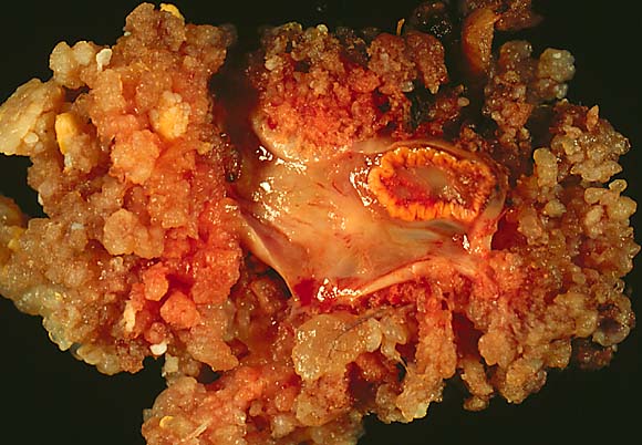 Ovarian surface papillary serous tumor of low malignant potential.
 -
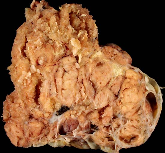 Serous cystadenocarcinoma of the ovary.


References
- ↑ Bradshaw, Karen D.; Schorge, John O.; Schaffer, Joseph; Lisa M. Halvorson; Hoffman, Barbara G. (2008). Williams' Gynecology. McGraw-Hill Professional. ISBN 0-07-147257-6.
- ↑ Kosary, Carol L. (2007). "Chapter 16: Cancers of the Ovary" (PDF). In Baguio, RNL; Young, JL; Keel, GE; Eisner, MP; Lin, YD; Horner, M-J. SEER Survival Monograph: Cancer Survival Among Adults: US SEER Program, 1988-2001, Patient and Tumor Characteristics. SEER Program. NIH Pub. No. 07-6215. Bethesda, MD: National Cancer Institute. pp. 133–144.
- ↑ 3.00 3.01 3.02 3.03 3.04 3.05 3.06 3.07 3.08 3.09 3.10 3.11 Surface epithelial-stromal tumor. Wikipedia. https://en.wikipedia.org/wiki/Surface_epithelial-stromal_tumor Accessed on April 18, 2016
- ↑ 4.0 4.1 4.2 4.3 4.4 4.5 4.6 4.7 Bell DA (1991). "Ovarian surface epithelial-stromal tumors". Hum. Pathol. 22 (8): 750–62. PMID 1869263.
- ↑ Kumar: Robbins and Cotran: Pathologic Basis of Disease, 7th ed.
- ↑ 6.0 6.1 Howlader N, Noone AM, Krapcho M, Garshell J, Miller D, Altekruse SF, Kosary CL, Yu M, Ruhl J, Tatalovich Z,Mariotto A, Lewis DR, Chen HS, Feuer EJ, Cronin KA (eds). SEER Cancer Statistics Review, 1975-2011, National Cancer Institute. Bethesda, MD, http://seer.cancer.gov/csr/1975_2011/, based on November 2013 SEER data submission, posted to the SEER web site, April 2014.