Primary amoebic meningoencephalitis pathophysiology
|
Primary amoebic meningoencephalitis Microchapters |
|
Differentiating Primary Amoebic Meningoencephalitis from other Diseases |
|---|
|
Diagnosis |
|
Treatment |
|
Case Studies |
|
Primary amoebic meningoencephalitis pathophysiology On the Web |
|
American Roentgen Ray Society Images of Primary amoebic meningoencephalitis pathophysiology |
|
Primary amoebic meningoencephalitis pathophysiology in the news |
|
Blogs on Primary amoebic meningoencephalitis pathophysiology |
|
Directions to Hospitals Treating Primary amoebic meningoencephalitis |
|
Risk calculators and risk factors for Primary amoebic meningoencephalitis pathophysiology |
Editor-In-Chief: C. Michael Gibson, M.S., M.D. [1]; Associate Editor(s)-in-Chief: Hardik Patel, M.D.
Overview
Naegleria fowleri is a heat-loving (thermophilic), free-living ameba (single-celled microbe), commonly found around the world in warm fresh water (like lakes, rivers, and hot springs) and soil that causes acute, fulminant hemorrhagic meningoencephalitis (primary amebic meningoencephalitis – PAM). Naegleria fowleri is the only species of Naegleria known to infect people. Most of the time, Naegleria fowleri lives in freshwater habitats by feeding on bacteria. However, in rare instances, the ameba can infect humans by entering the nose during water-related activities. Once in the nose, the ameba travels to the brain and causes a severe brain infection, primary amoebic meningoencephalitis (PAM), which is usually fatal.
Pathophysiology
Naegleria fowleri propagates in warm, stagnant bodies of freshwater (typically during the summer months), and enters the central nervous system after insufflation of infected water by attaching itself to the olfactory nerve.[1] It then migrates through the cribiform plate and into the olfactory bulbs of the forebrain,[2] where it multiplies itself greatly by feeding on nerve tissue. During this stage, occurring approximately 3–7 days post-infection, the typical symptoms are parosmia, rapidly progressing to anosmia (with resultant ageusia) as the nerve cells of the olfactory bulbs are consumed and replaced with necrotic lesions. After the organisms have multiplied and largely consumed the olfactory bulbs, the infection rapidly spreads through the mitral cell axons to the rest of the cerebrum, resulting in onset of symptoms.
Life Cycle

Naegleria fowleri has 3 stages in its life cycle: ameboid trophozoites, flagellates, and cysts. The only infective stage of the ameba is the ameboid trophozoite. Trophozoites are 10-35 µm long with a granular appearance and a single nucleus. The trophozoites replicate by binary division during which the nuclear membrane remains intact (a process called promitosis). Trophozoites infect humans or animals by penetrating the nasal tissue and migrating to the brain via the olfactory nerves causing primary amebic meningoencephalitis (PAM). Trophozoites can turn into a temporary, non-feeding, flagellated stage (10-16 µm in length) when stimulated by adverse environmental changes such as a reduced food source. They revert back to the trophozoite stage when favorable conditions return. Naegleria fowleri trophozoites are found in cerebrospinal fluid (CSF) and tissue, while flagellated forms are occasionally found in CSF. Cysts are not seen in brain tissue. If the environment is not conducive to continued feeding and growth (like cold temperatures, food becomes scarce) the ameba or flagellate will form a cyst. The cyst form is spherical and about 7-15 µm in diameter. It has a smooth, single-layered wall with a single nucleus. Cysts are environmentally resistant in order to increase the chances of survival until better environmental conditions occur.
 |
 |
 |
Copyleft images obtained courtesy of http://www.cdc.gov/parasites/naegleria/pathogen.html
Gross Pathology Images


Copyleft images obtained courtesy of http://www.cdc.gov/parasites/naegleria/naegleria-fowleri-images.html
Microscopic Pathology Images
-
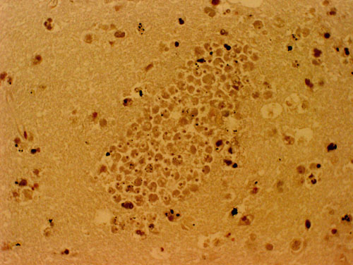 A section of the brain stained with hematoxylin and eosin, of a PAM patient showing a large cluster of Naegleria fowleri trophozoites. Cysts are not seen.
 -
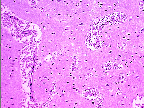 A section of the brain, stained with hematoxylin and eosin, of a PAM patient showing large clusters of Naegleria fowleri trophozoites. Cysts are not seen.


Copyleft images obtained courtesy of http://www.cdc.gov/parasites/naegleria/naegleria-fowleri-images.html
References
- ↑ Centers for Disease Control and Prevention (CDC) (2008). "Primary amebic meningoencephalitis—Arizona, Florida, and Texas, 2007". MMWR Morb. Mortal. Wkly. Rep. 57 (21): 573–7. PMID 18509301. Unknown parameter
|month=ignored (help) - ↑ Cervantes-Sandoval I, Serrano-Luna Jde J, García-Latorre E, Tsutsumi V, Shibayama M (2008). "Characterization of brain inflammation during primary amoebic meningoencephalitis". Parasitol. Int. 57 (3): 307–13. doi:10.1016/j.parint.2008.01.006. PMID 18374627. Unknown parameter
|month=ignored (help)