Mucoepidermoid carcinoma echocardiography or ultrasound
|
Mucoepidermoid carcinoma Microchapters |
|
Differentiating Mucoepidermoid Carcinoma from other Diseases |
|---|
|
Diagnosis |
|
Treatment |
|
Case Studies |
|
Mucoepidermoid carcinoma echocardiography or ultrasound On the Web |
|
American Roentgen Ray Society Images of Mucoepidermoid carcinoma echocardiography or ultrasound |
|
FDA on Mucoepidermoid carcinoma echocardiography or ultrasound |
|
CDC on Mucoepidermoid carcinoma echocardiography or ultrasound |
|
Mucoepidermoid carcinoma echocardiography or ultrasound in the news |
|
Blogs on Mucoepidermoid carcinoma echocardiography or ultrasound |
|
Risk calculators and risk factors for Mucoepidermoid carcinoma echocardiography or ultrasound |
Editor-In-Chief: C. Michael Gibson, M.S., M.D. [1]Associate Editor(s)-in-Chief: Badria Munir M.B.B.S.[2] , Maria Fernanda Villarreal, M.D. [3]
Overview
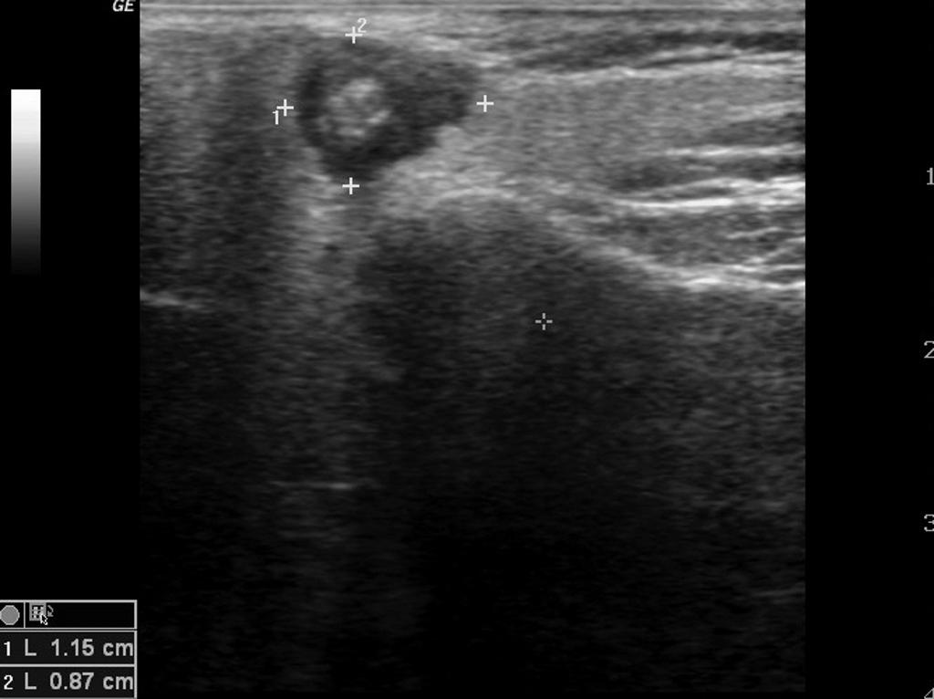
On ultrasound, characteristic findings of mucoepidermoid carcinoma include: a well-circumscribed hypoechoic lesion with a partial or completely cystic appearance, in contrast to the relatively hyperechoeic normal parotid gland.
 |
Ultrasound
- Ultrasound findings associated with mucoepidermoid carcinoma include:[1]
- It can determine location, homogeneity or heterogeneity, shape, vascularity, and margins of salivary tumors in the periauricular, buccal, and submandibular area.
- Ultrasonography may be reveal the type of tumor, in addition to that, new ultrasonographic contrast mediums can demonstrate the vascularity of the tumor before surgery.[2][3]
- Well-circumscribed hypoechoic lesion
- Partial or completely cystic appearance
References
- ↑ Mucoepidermoid carcinoma. Radiopedia. Dr Frank Gailliard. http://radiopaedia.org/articles/mucoepidermoid-carcinoma-of-salivary-glands Accessed on February 17, 2016
- ↑ Rong X, Zhu Q, Ji H, Li J, Huang H (December 2014). "Differentiation of pleomorphic adenoma and Warthin's tumor of the parotid gland: ultrasonographic features". Acta Radiol. 55 (10): 1203–9. doi:10.1177/0284185113515865. PMID 24324278.
- ↑ Yuan WH, Hsu HC, Chou YH, Hsueh HC, Tseng TK, Tiu CM (2009). "Gray-scale and color Doppler ultrasonographic features of pleomorphic adenoma and Warthin's tumor in major salivary glands". Clin Imaging. 33 (5): 348–53. doi:10.1016/j.clinimag.2008.12.004. PMID 19712813.