Inferior pharyngeal constrictor muscle
Template:Muscle infobox The Inferior pharyngeal constrictor, the thickest of the three constrictors, arises from the sides of the cricoid and thyroid cartilage. Similarly to the superior and middle pharyngeal constrictor muscles, it is innervated by the vagus nerve (cranial nerve X).
Origin and insertion
The components arising from the cricoid and thyroid cartilages are also known as cricopharyngeus and thyropharyngeus respectively. [1]
- From the cricoid cartilage it arises in the interval between the Cricothyreoideus in front, and the articular facet for the inferior cornu of the thyroid cartilage behind.
- On the thyroid cartilage it arises from the oblique line on the side of the lamina, from the surface behind this nearly as far as the posterior border and from the inferior cornu.
From these origins the fibers spread backward and medialward to be inserted with the muscle of the opposite side into the fibrous raphé in the posterior median line of the pharynx.
The inferior fibers are horizontal and continuous with the circular fibers of the esophagus; the rest ascend, increasing in obliquity, and overlap the Constrictor medius.
Action
As soon as the bolus of food is received in the pharynx, the elevator muscles relax, the pharynx descends, and the constrictores contract upon the bolus, and convey it downward into the esophagus.
Role in human disease
Uncoordinated contraction, and/or spasm and/or impaired relaxation of this muscle are currently considered the main factors in development of a Zenker's diverticulum.
Motor incoordination of the cricopharyngeus can cause difficulty swallowing. Severe spasm can cause pain during swallowing(Odynophagia).
See also
Additional images
-
 Muscles of the neck. Lateral view.
 -
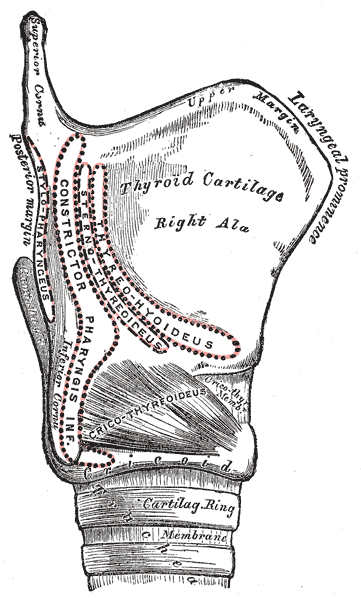 Side view of the larynx, showing muscular attachments.


References
External links
Template:Gray's Template:Muscles of head
de:Musculus constrictor pharyngis inferior hu:Alsó garat-összeszorító izom sr:Мишић доњи констриктор ждрела sh:Mišić donji konstriktor ždrela Template:WH Template:WikiDoc Sources